Surgeon/Restorative dentist:
Dr. Leonardo Targetti – Florence, Italy
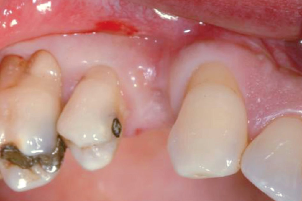
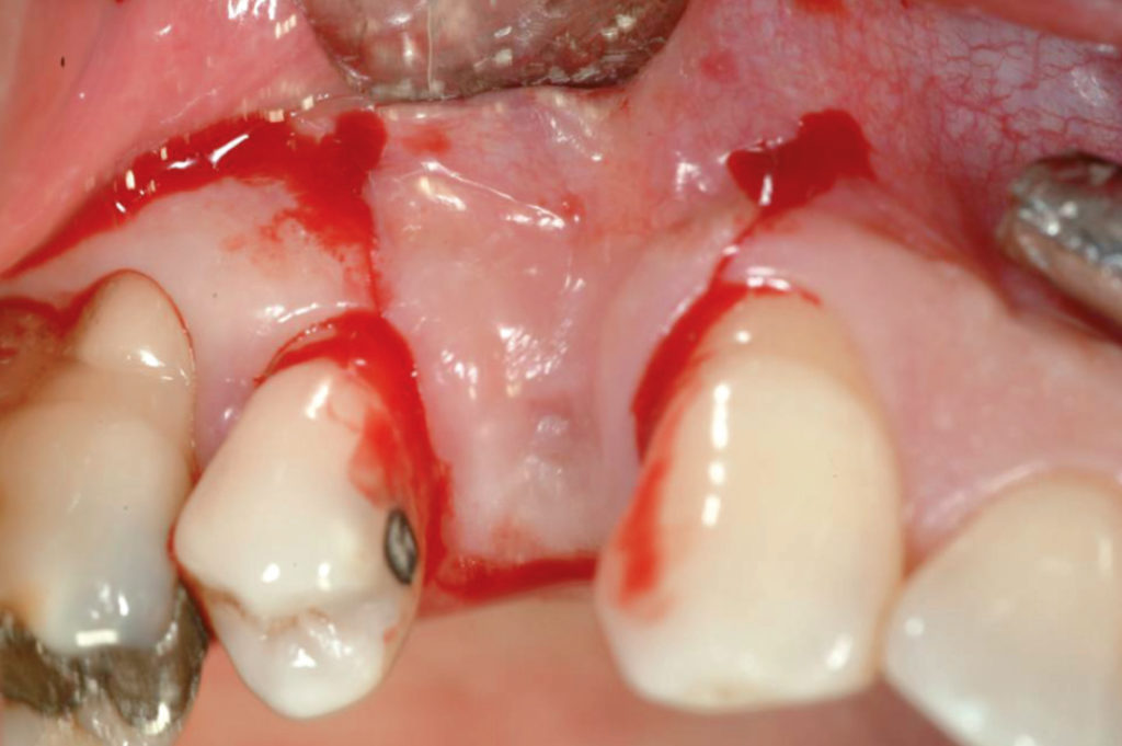
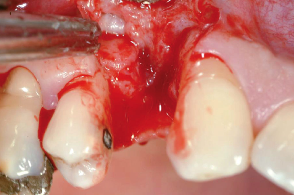
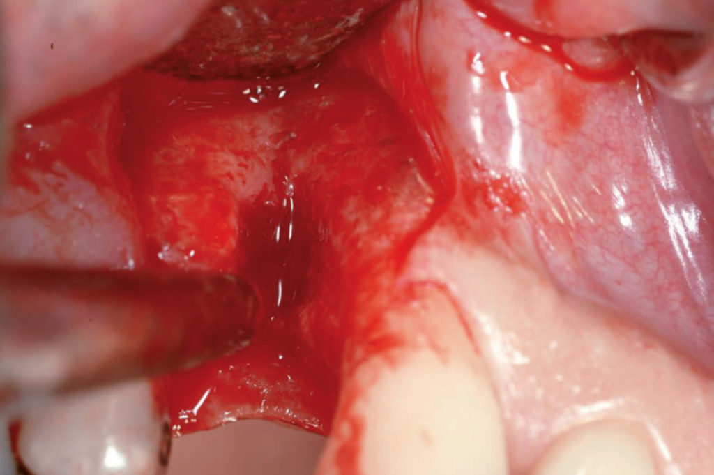
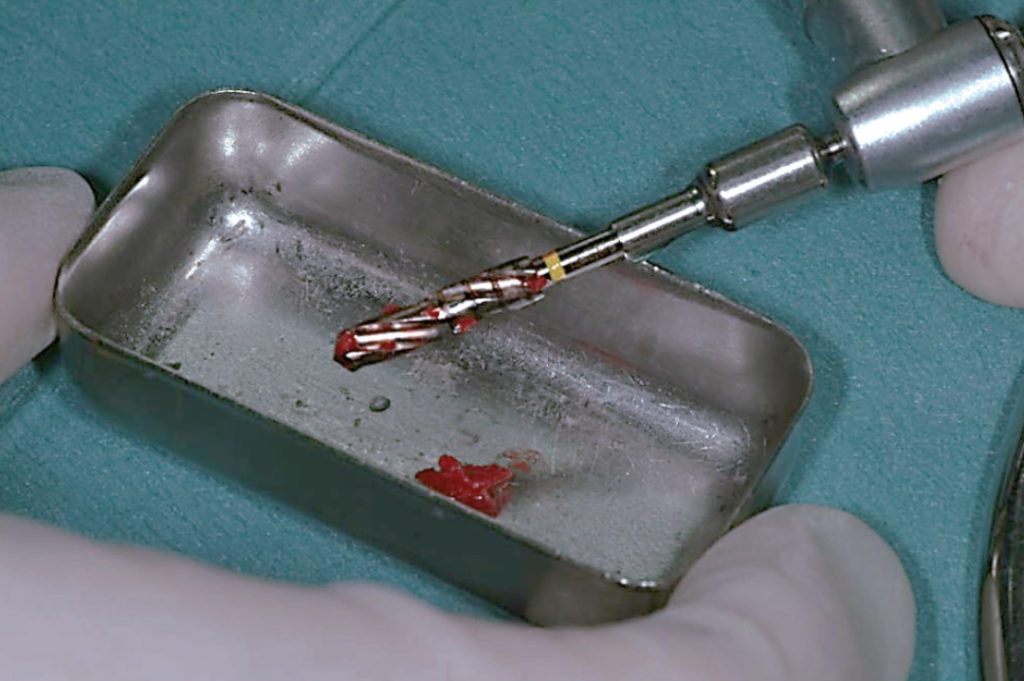
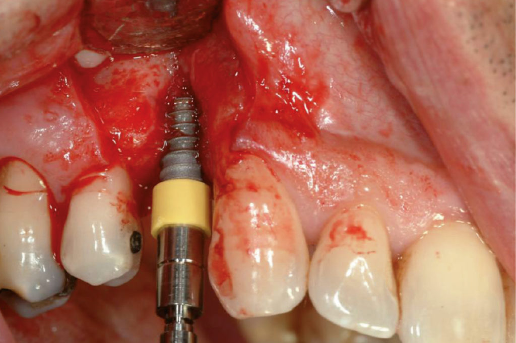
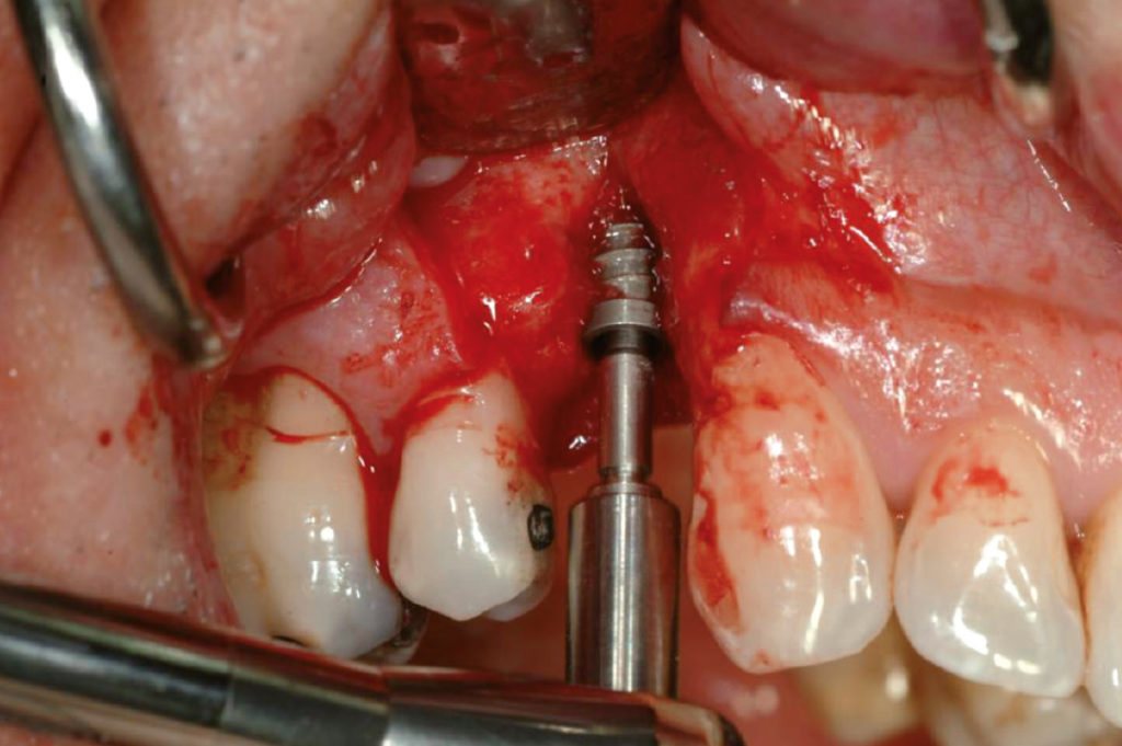
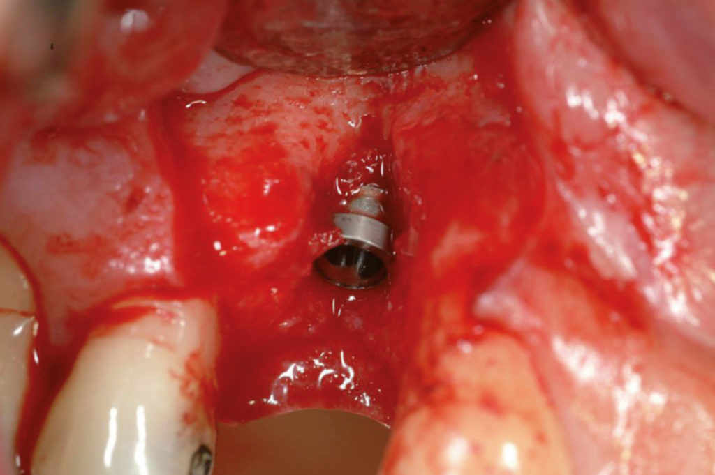
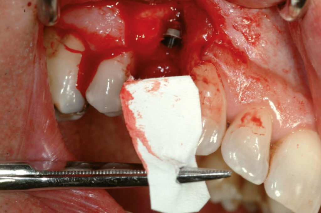
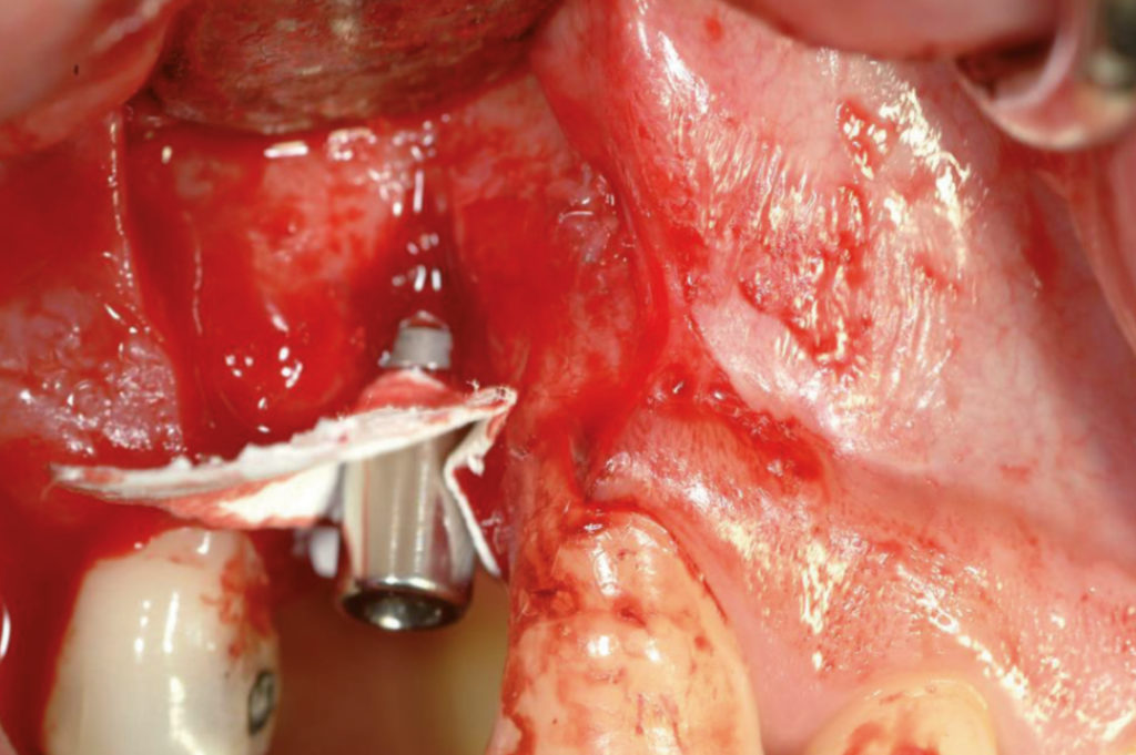
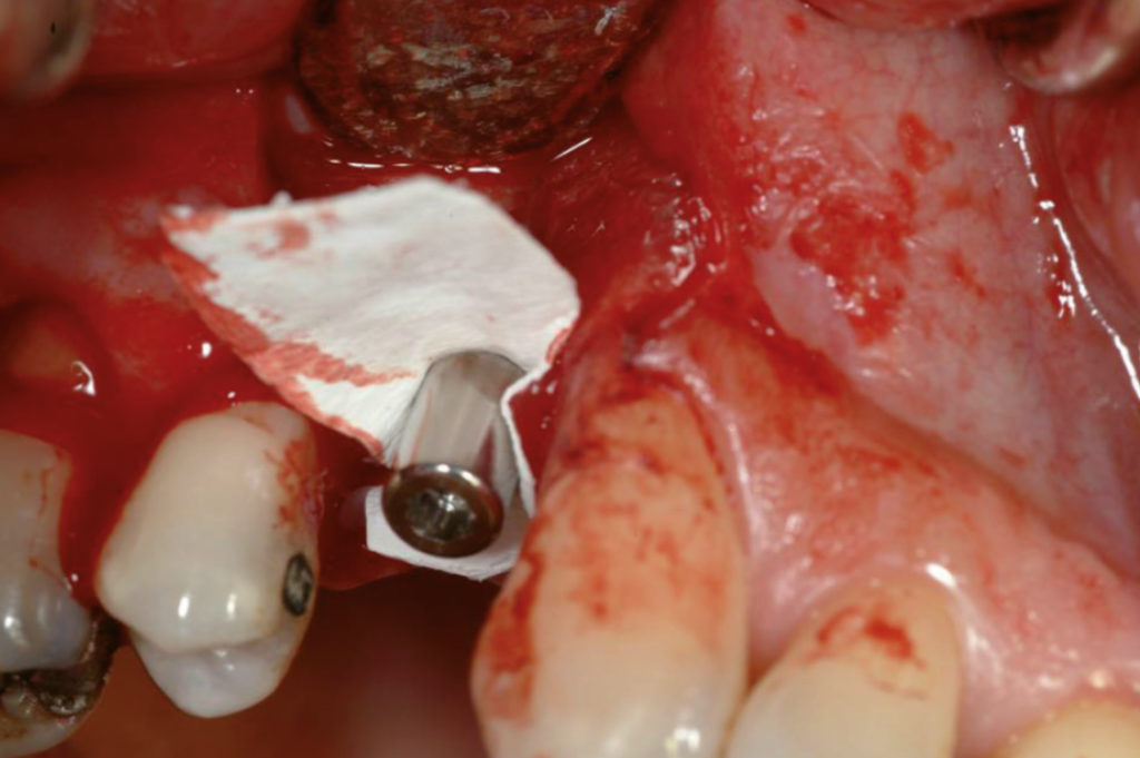
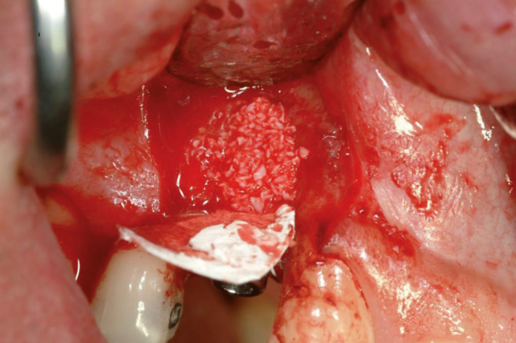
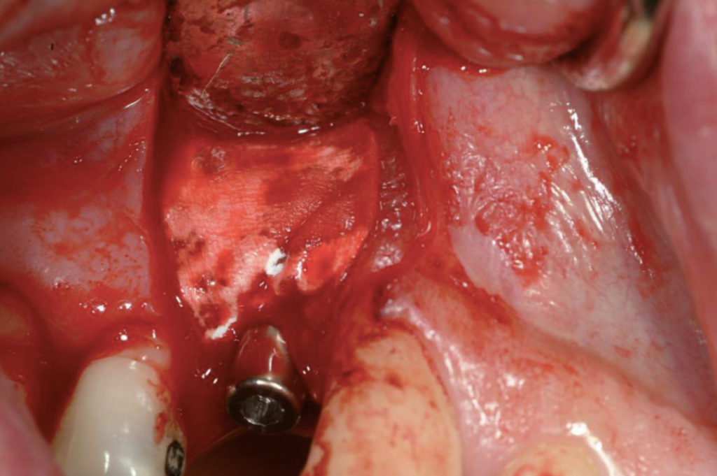
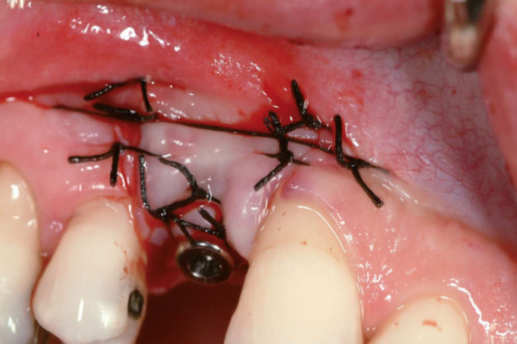
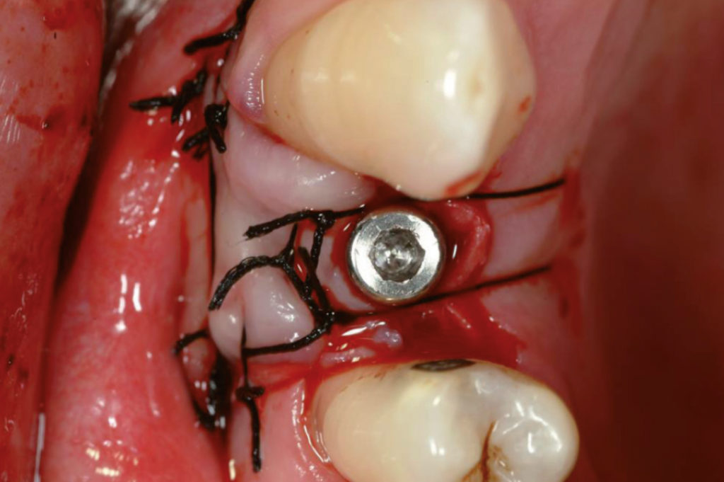
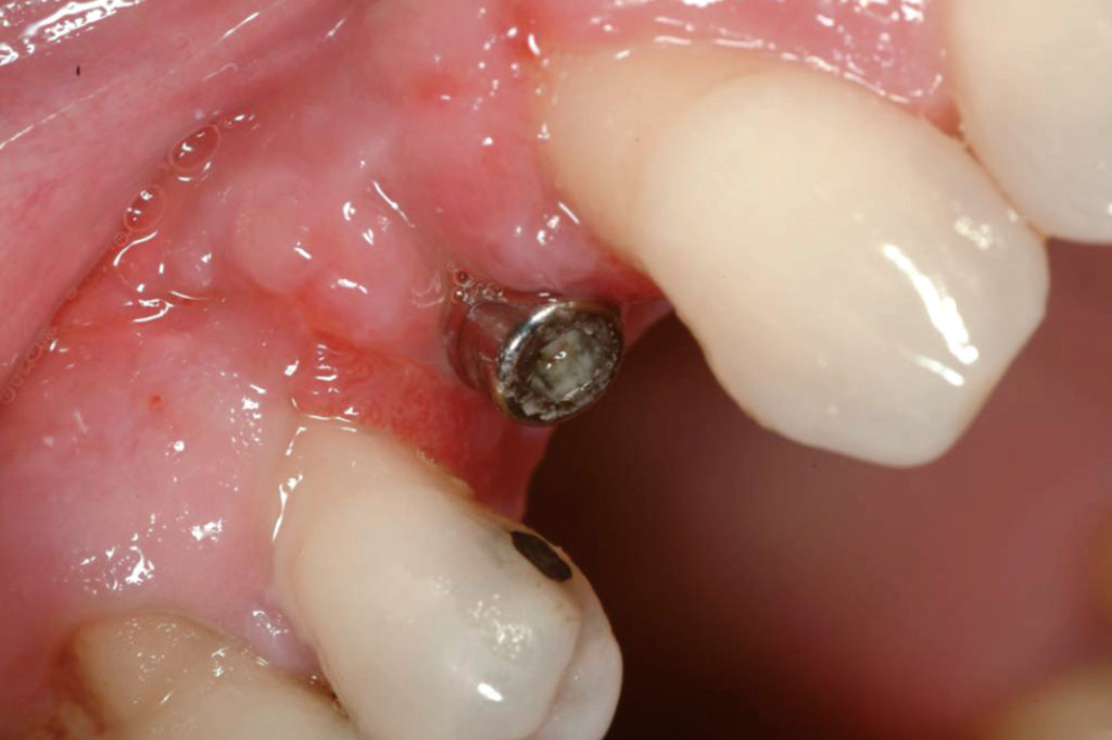
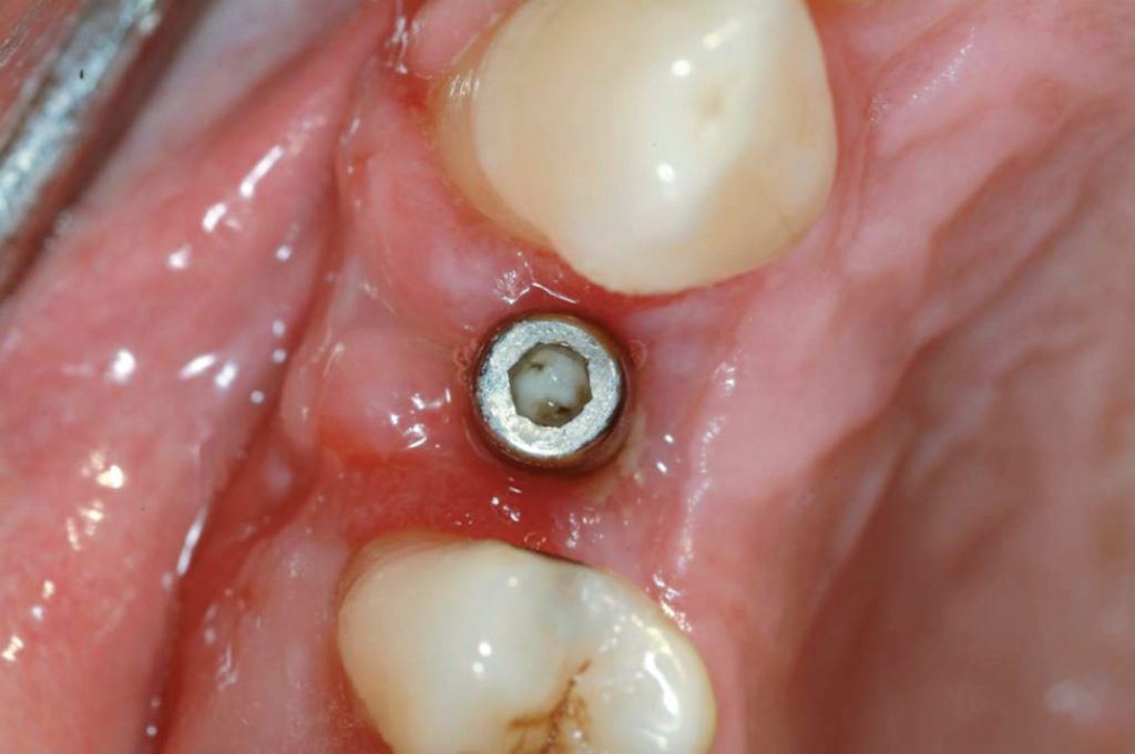
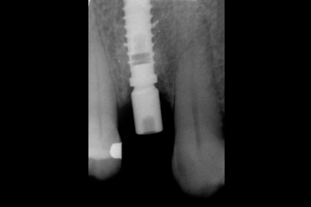
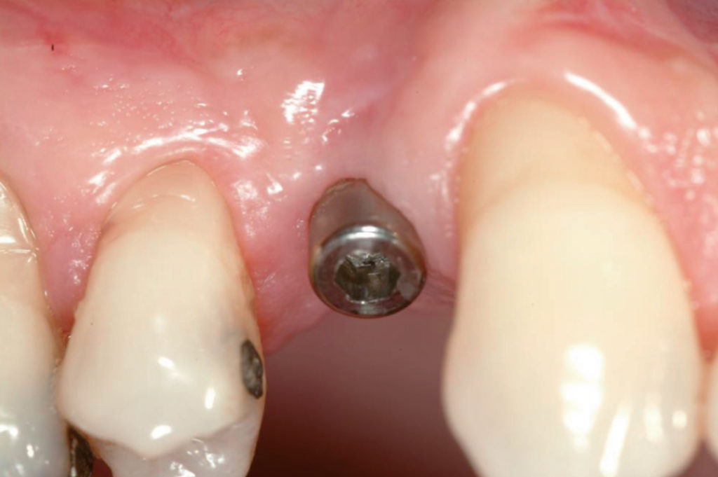
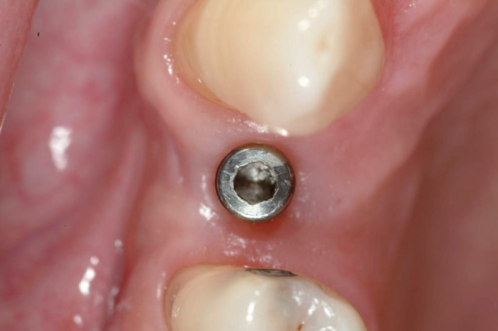
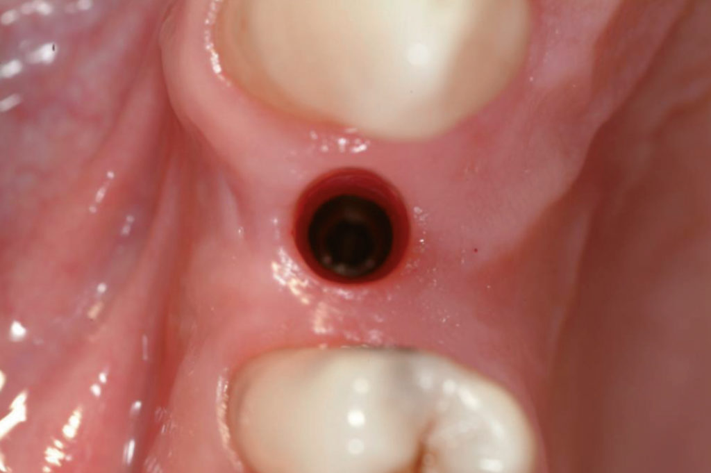
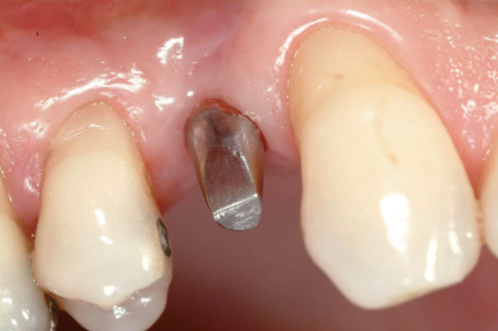
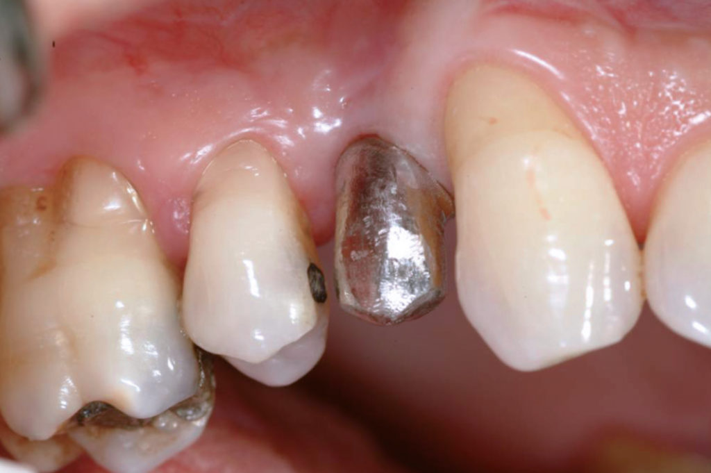
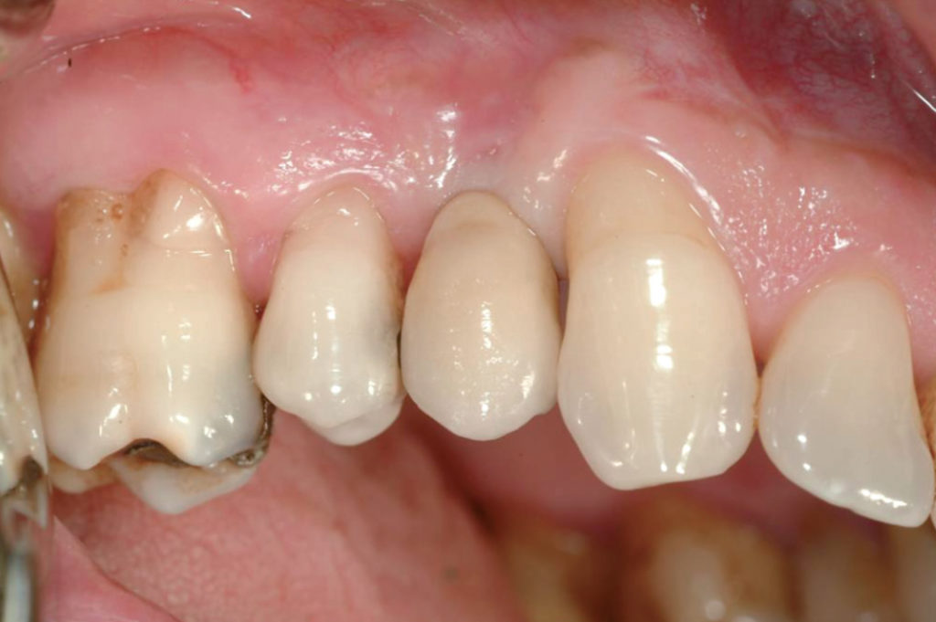
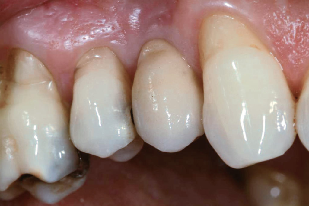
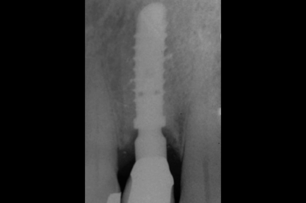
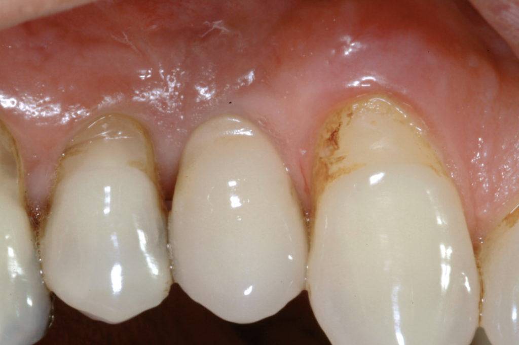
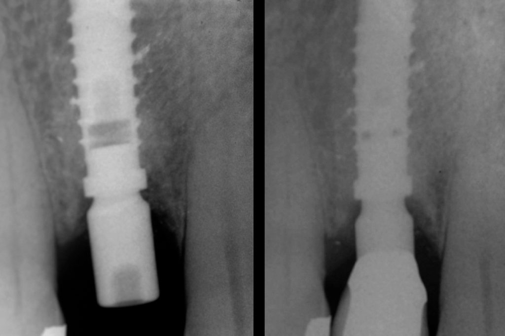
This case report demonstrates a procedure for restoring a missing maxillary first premolar with a severe buccal bone plate defect of a 55-year-old male patient. In order to expose the bone defect a GBR incision was performed and a full-thickness mucoperiosteal flap was raised. After thorough surgical debridement of the defect a 4.1 x 14 mm XCN Classix implant was placed about 3 mm subcrestally. A small hole was made in a collagen membrane in order to stabilize the membrane with a healing cap for a one-stage surgical procedure. The bone defect was filled with a mixture of autologous bone, harvested from the drills during preparation of the implant site, and a bone graft substitute. After six months of healing an implant level impression was taken and a cement-retained metal-ceramic crown was fabricated. In May 2016, twelve years after crown delivery, clinical and radiographic examination was performed. The clinical picture shows stable peri-implant soft tissue levels and aesthetics. The comparison of X-ray follow-up, at six months and at twelve years after crown delivery, demonstrates no crestal bone loss and reveals a gain in peri-implant crestal bone height.

Pre-operative clinical view GBR incision Full-thickness mucoperiosteal flap View of severe bone defect after surgical debridement Bone chips collected during implant bed preparation by low speed drilling without irrigation Insertion of 4.1 x 14 mm XCN Classix implant Implant insertion in a subcrestal position View of implant in its final position, about 3 mm subcrestally Creation of a small hole within the membrane Placement of a healing cap with a height of 7 mm in order to stabilize the membrane Placement of a healing cap with a height of 7 mm in order to stabilize the membrane Filling of the bone defect with a mixture of autologous bone and bone graft substitute View of the bone graft covered by the membrane Wound closure with interrupted sutures View of the healing cap after suturing Fifteen days post-operative clinical view Fifteen days post-operative clinical view Six months post-operative X-ray Six months post-operative clinical view Six months post-operative clinical view View of peri-implant soft tissues after removal of the healing cap Try-in of the prepared abutment Try-in of the metal structure Delivery of the cement-retained metal-ceramic crown Clinical situation four years after crown delivery Twelve-year X-ray follow-up Twelve-year clinical follow-up. Note stable peri-implant soft tissue levels and aesthetics Comparison of six-month and twelve-year X-ray follow-up. Note the gain in peri-implant crestal bone height
Laboratory:
Picchi, Perugi and Santoni, Danilo Petroni & C. – Florence, Italy