Cirujano/ Dentista restaurador:
Dr. Leonardo Targetti – Florencia, Italia
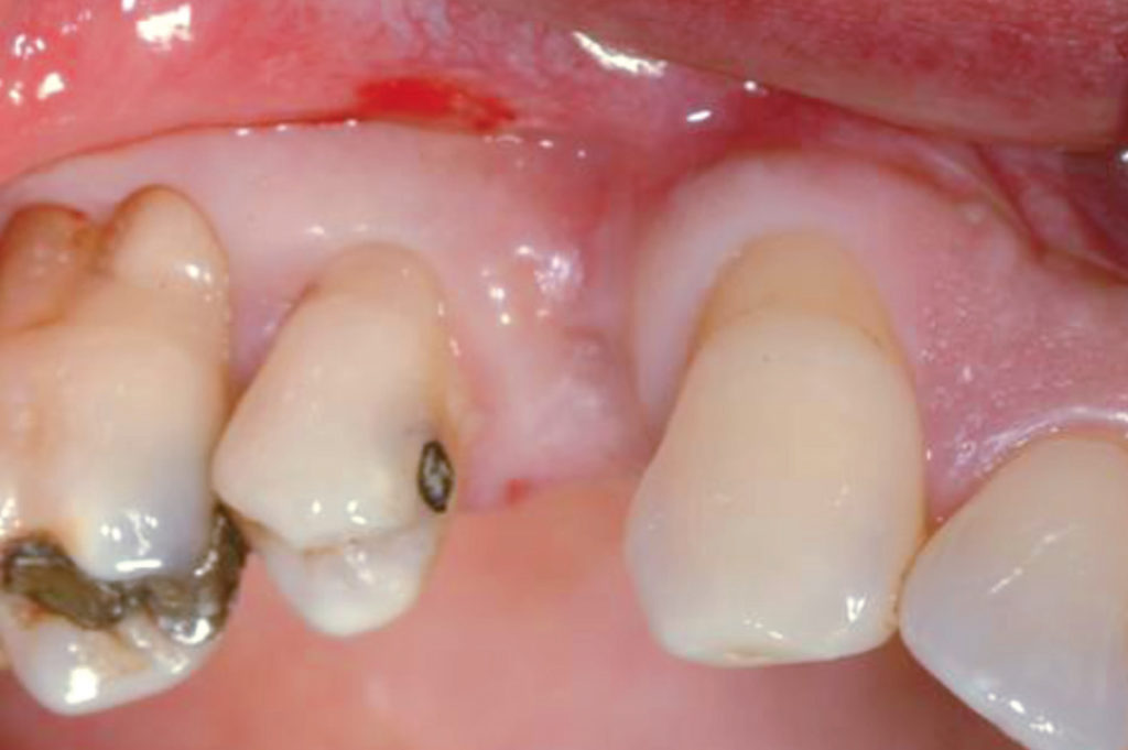
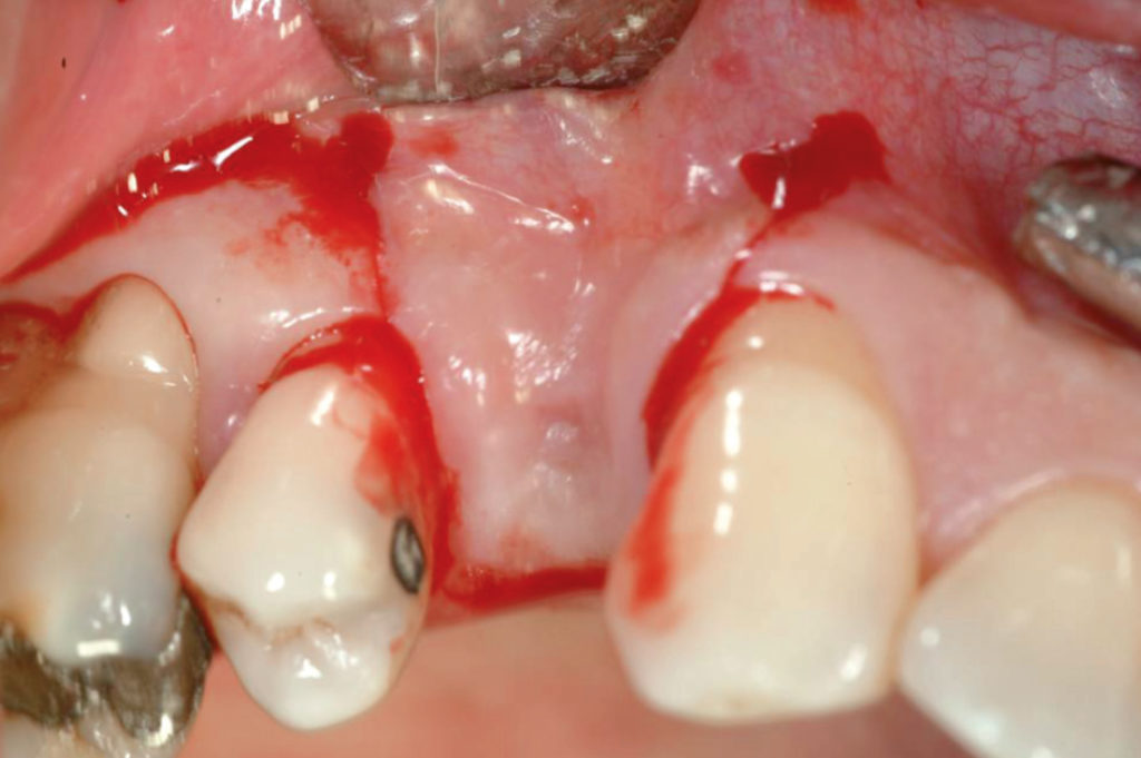
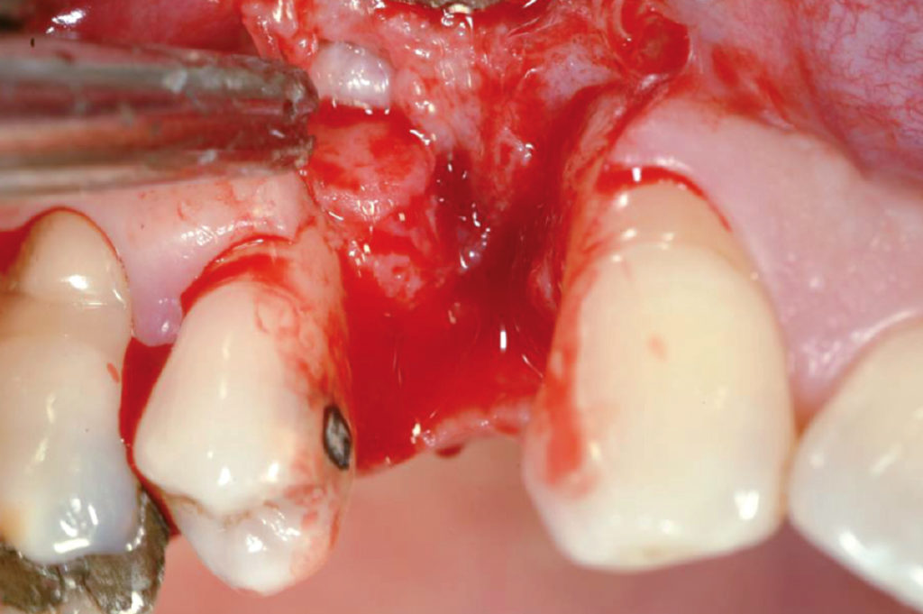
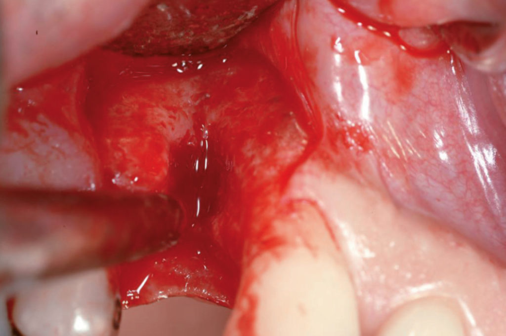
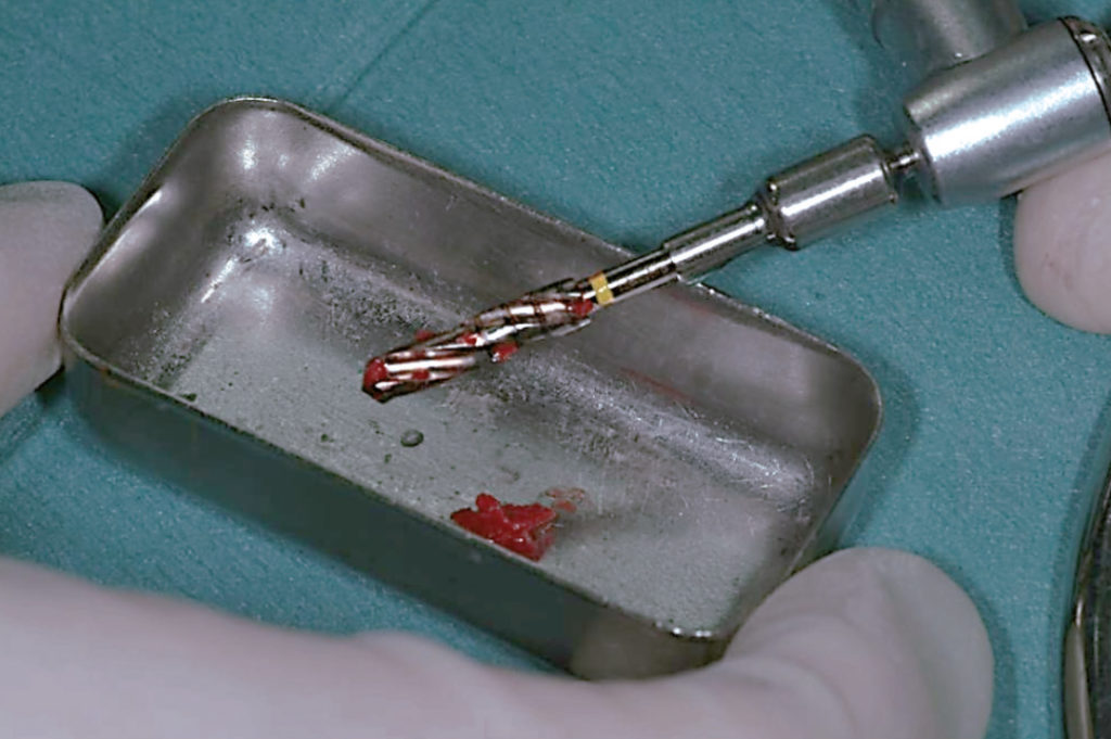
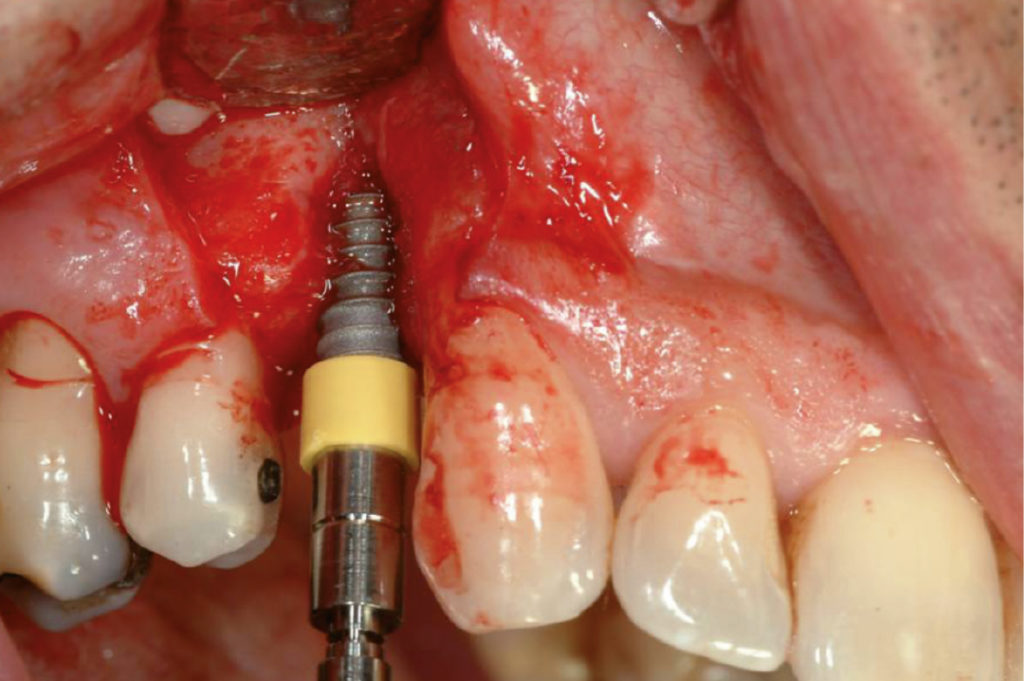
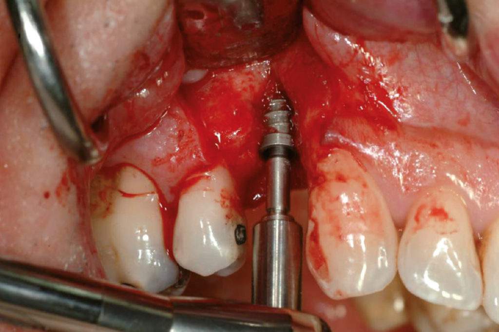
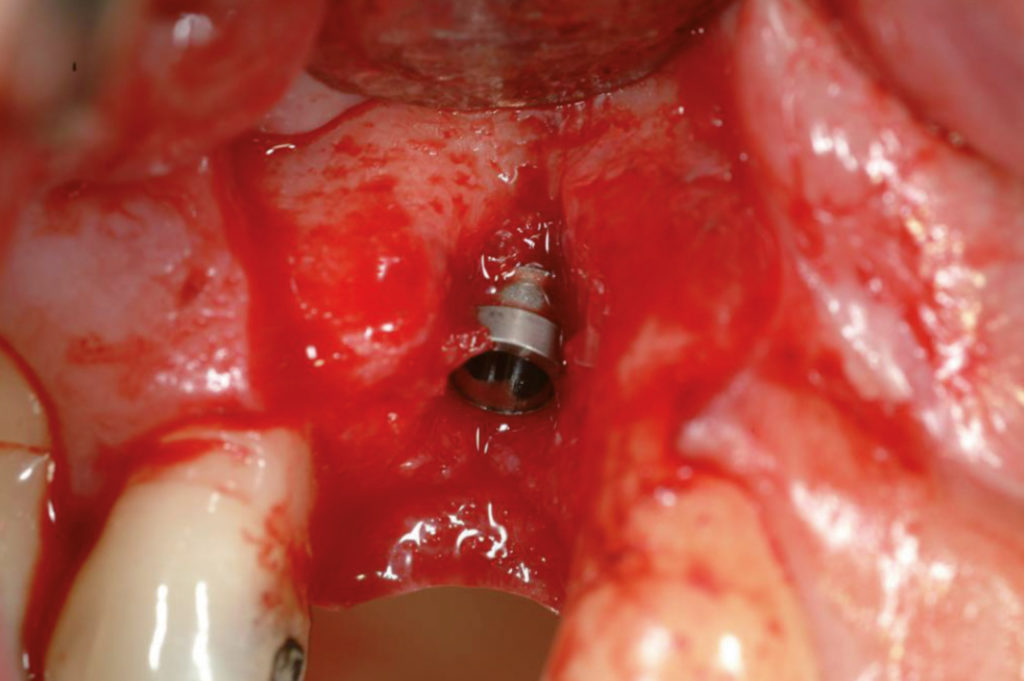
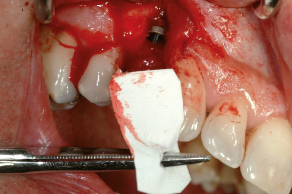
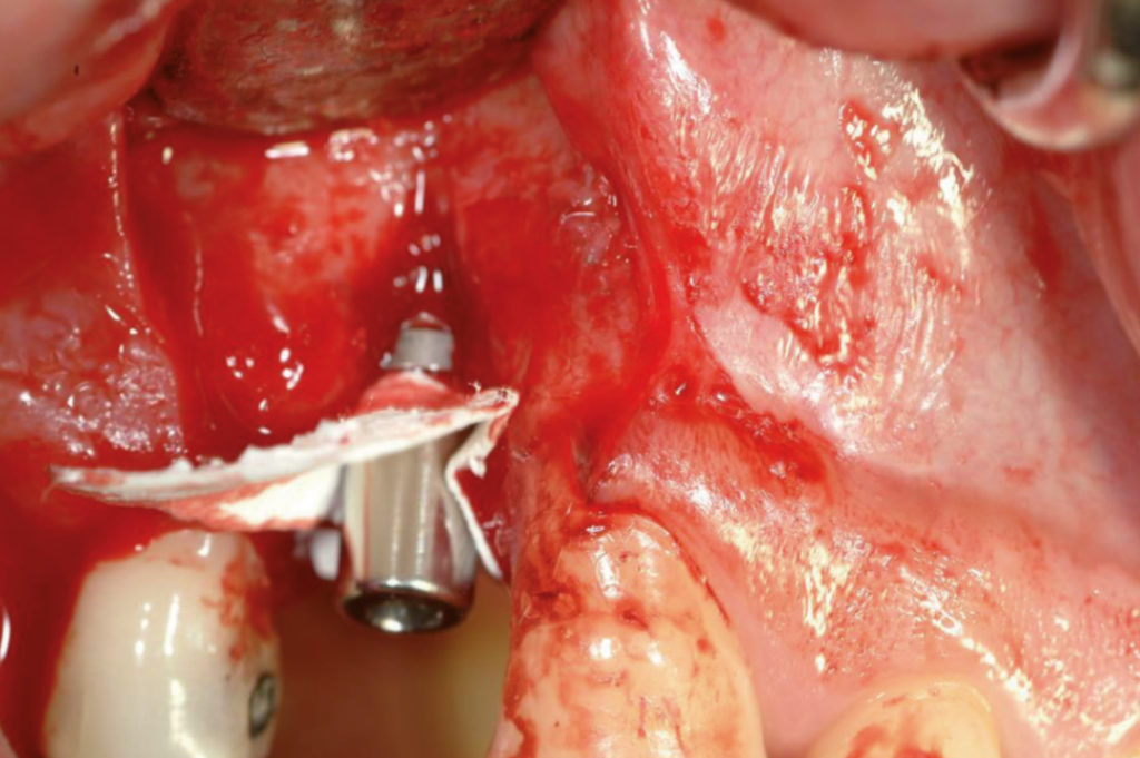
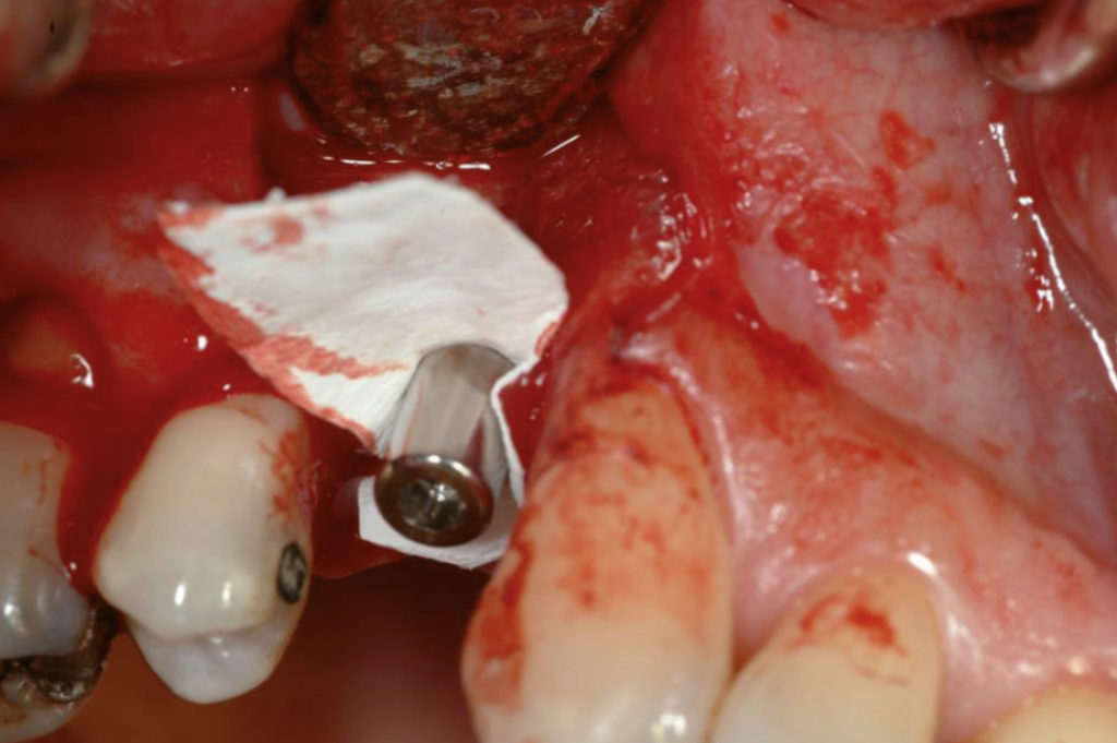
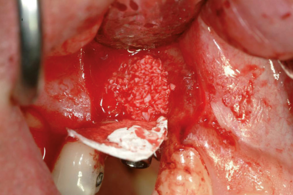
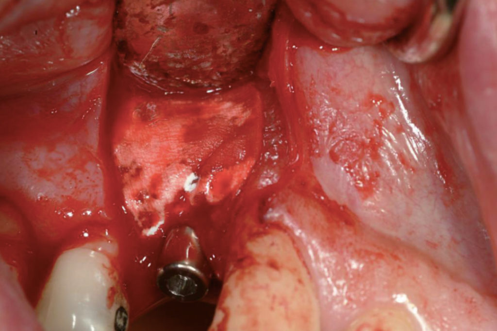
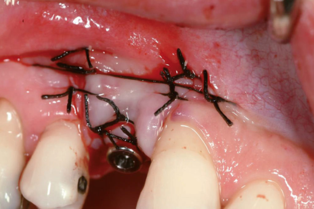
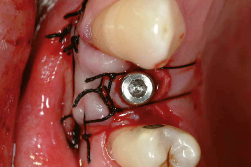
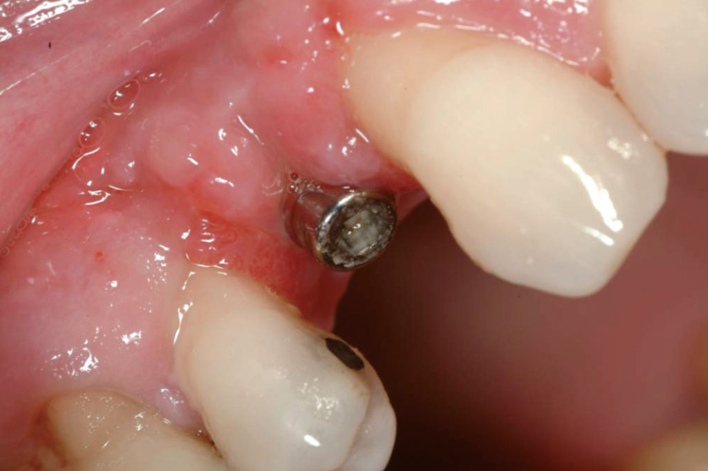
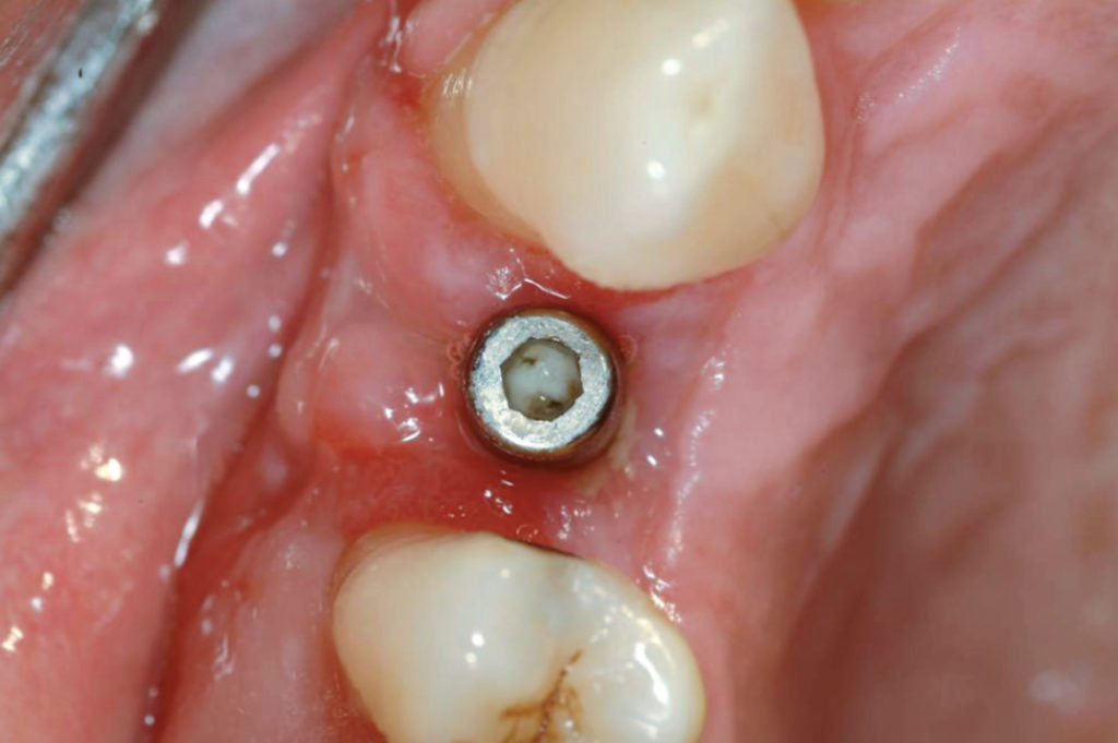
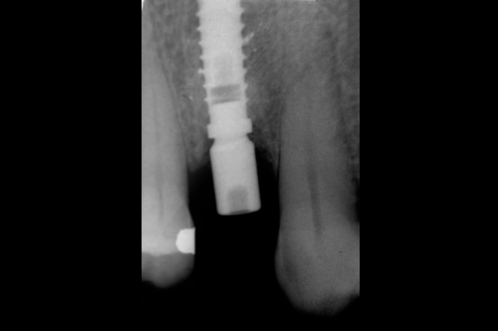
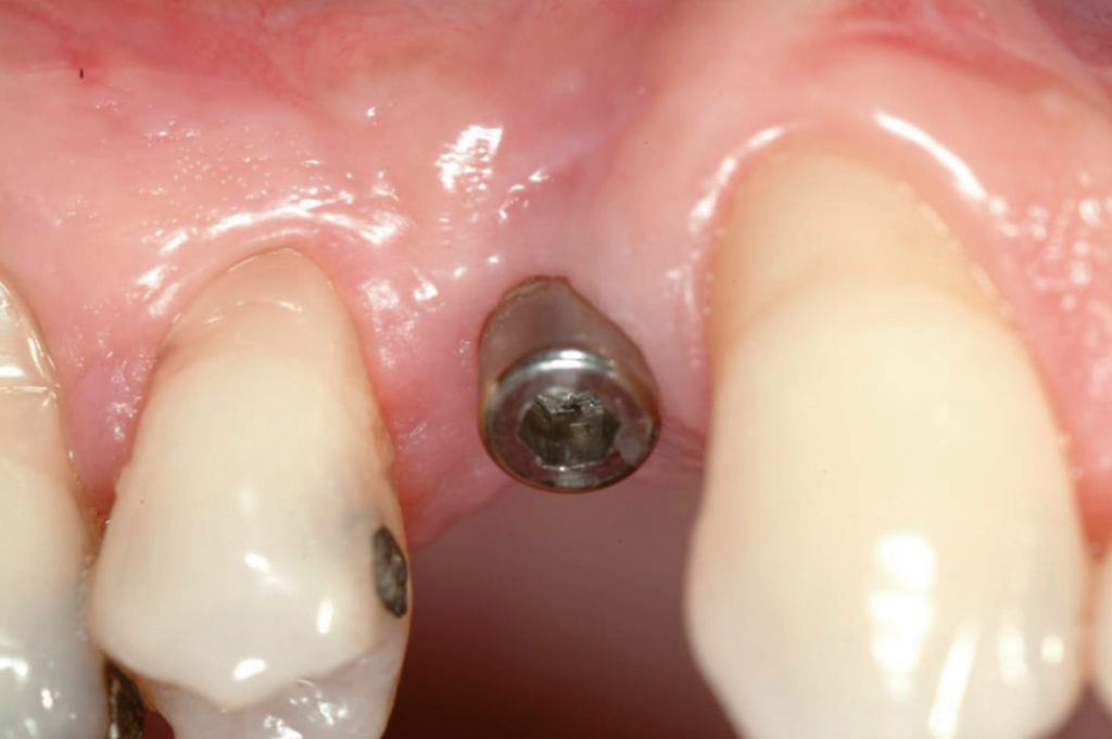
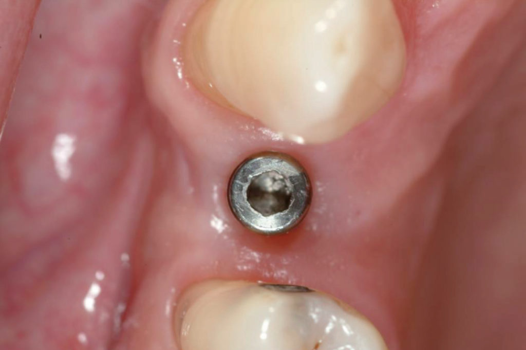
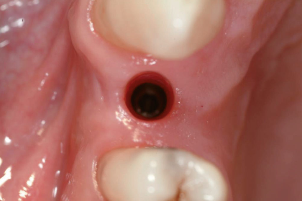
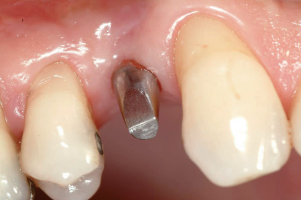
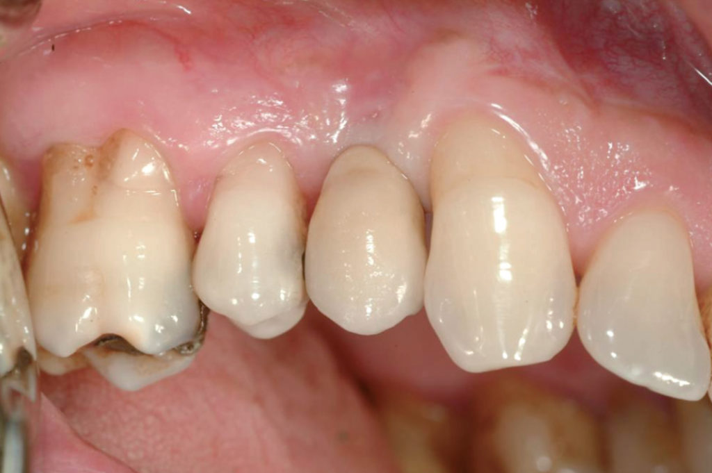
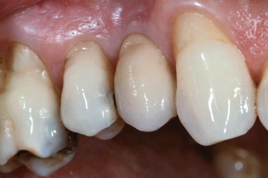
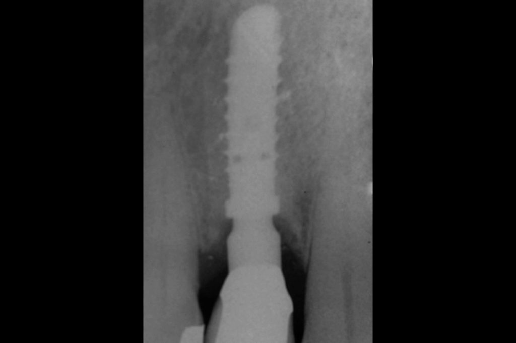
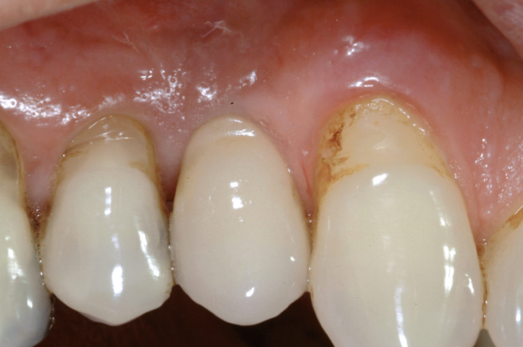
This case report demonstrates a procedure for restoring a missing maxillary first premolar with a severe buccal bone plate defect of a 55-year-old male patient. In order to expose the bone defect a GBR incision was performed and a full-thickness mucoperiosteal flap was raised. After thorough surgical debridement of the defect a 4.1 x 14 mm XCN Classix implant was placed about 3 mm subcrestally. A small hole was made in a collagen membrane in order to stabilize the membrane with a healing cap for a one-stage surgical procedure. The bone defect was filled with a mixture of autologous bone, harvested from the drills during preparation of the implant site, and a bone graft substitute. After six months of healing an implant level impression was taken and a cement-retained metal-ceramic crown was fabricated. In May 2016, twelve years after crown delivery, clinical and radiographic examination was performed. The clinical picture shows stable peri-implant soft tissue levels and aesthetics. The comparison of X-ray follow-up, at six months and at twelve years after crown delivery, demonstrates no crestal bone loss and reveals a gain in peri-implant crestal bone height.

Vista clínica preoperatoria Incisión GBR Levantamiento del colgajo de espesor completo mucoperióstico Vista del fuerte defecto óseo después de desbridamiento quirúrgico Astillas de hueso recogidas durante la preparación del lecho del implante por medio de fresado a baja velocidad sin irrigación Colocación del implante XCN® Classix 4.1 x 14 mm Colocación del implante en posición subcrestal Vista del implante en su posición final, unos 3 mm subcrestalmente Creación de un pequeño opérculo dentro de la membrana Posicionamiento de un tapón de cicatrización de 7 mm de altura para estabilizar la membrana Tapón de cicatrización posicionado, gracias a su conexión cono Morse el tapón de cicatrización ofrece un sellado bacteriano perfecto Llenado del hueso con una mezcla de autólogo de médula ósea y sustituto de injerto óseo Vista del injerto cubierto por la membrana Cierre de la herida con suturas interrumpidas Vista del tapón de cicatrización después de la sutura Vista clínica postoperatoria después de quince días Vista clínica postoperatoria después de quince días Rx postoperatoria después de seis meses Vista clínica postoperatoria después de seis meses Vista clínica postoperatoria después de seis meses Vista de los tejidos blandos periimplantarios después de la remoción del tapón de cicatrización Prueba del pilar preparado Prueba de la estructura de metal Entrega de la corona cementada de metal-cerámica Situación clínica cinco años después de la entrega de la corona Rx de seguimiento a los 12 años. Obsérvese niveles periimplantarios de los tejidos blandos y estética estables Comparación de Rx de seguimiento a los seis meses y a los doce meses. Obsérvese la ganancia en la altura del hueso crestal periimplantario Comparación de Rx de seguimiento a los seis meses y a los doce meses. Obsérvese la ganancia en la altura del hueso crestal periimplantario


Laboratorio:
Picchi, Perugi y Santoni, Danilo Petroni & C. – Florencia, Italia