Surgeon/Restorative dentist:
Dr. Leonardo Palazzo – Gubbio (Perugia), Italy
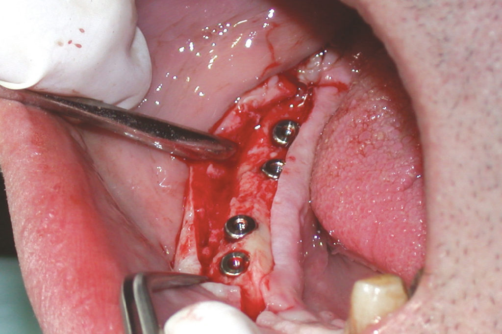
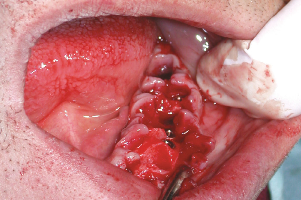
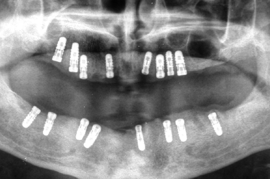
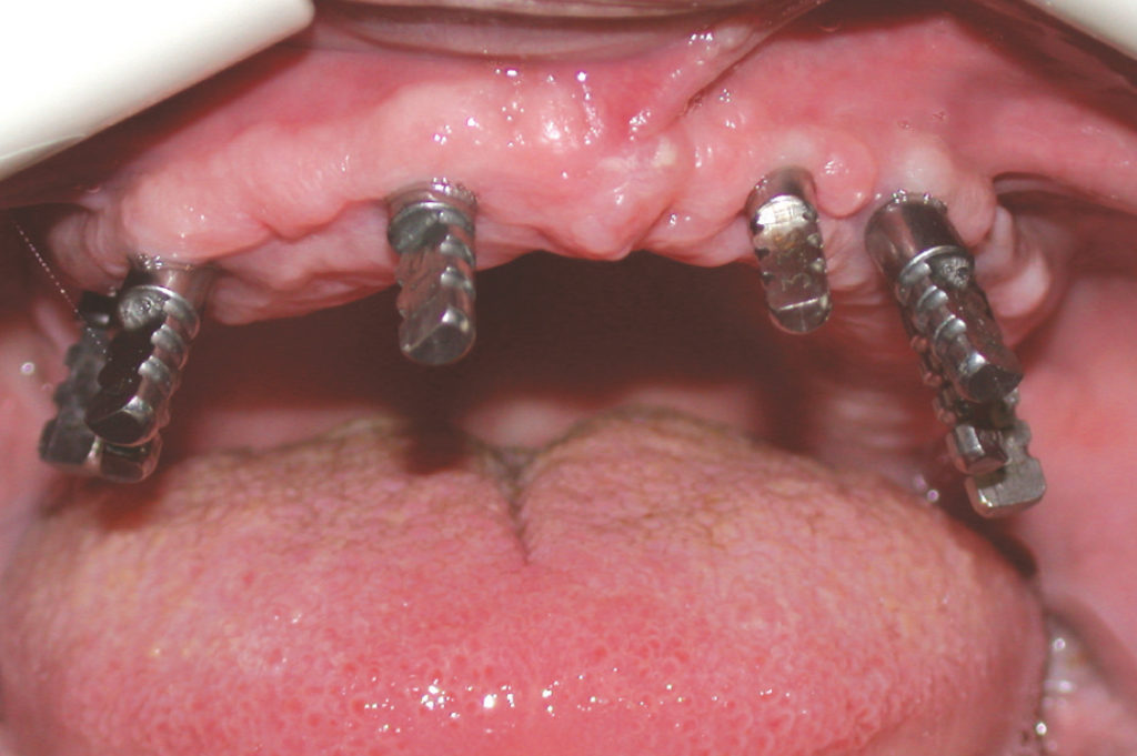
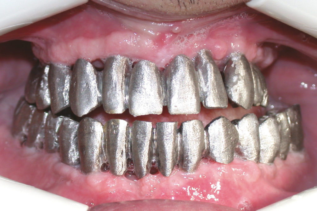
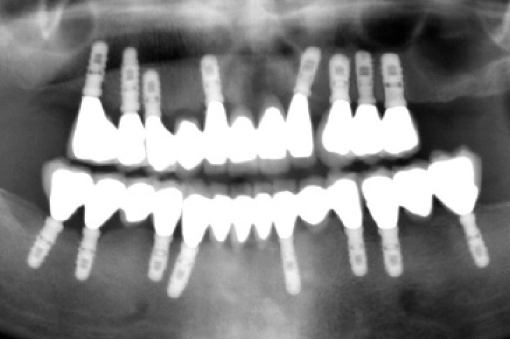
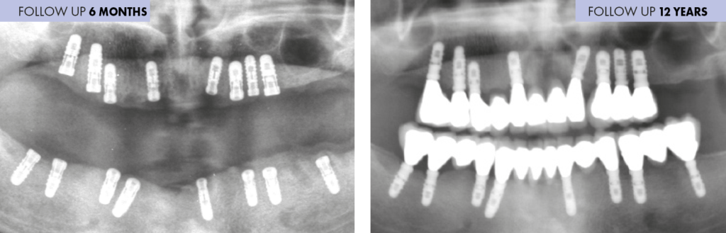
This case report illustrates the treatment of a 58-year-old male patient with a critical periodontal situation and numerous missing teeth in both jaws asking for implant supported fixed restorations. Clinical and radiographic examination revealed sufficient bone volume for the placement of implants in both jaws. After extraction of all residual teeth and removal of a maxillary cyst, eight XCN implants were placed in the upper jaw with a two-stage surgical procedure. During the same treatment session two-stage placement of further eight Leone implants in the lower jaw was performed. After six months of healing, the implants were uncovered, impressions were taken and cement-retained metal-ceramic prosthesis were fabricated. Panoramic radiograph and clinical pictures twelve years after delivery of the final prostheses show peri-implant bone stability and stable soft tissue margins.

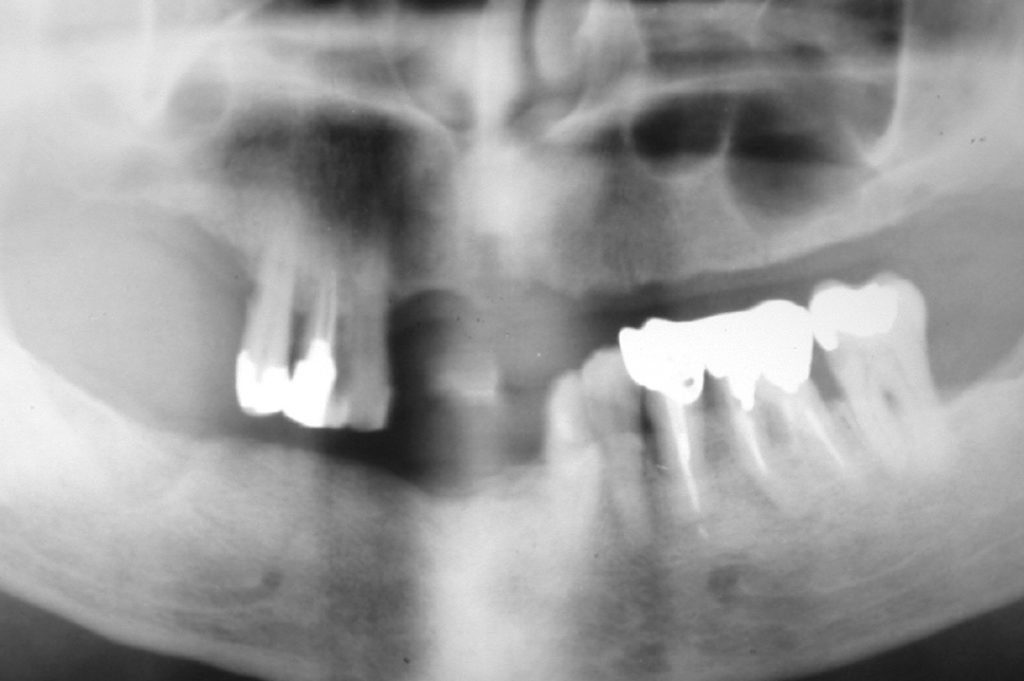
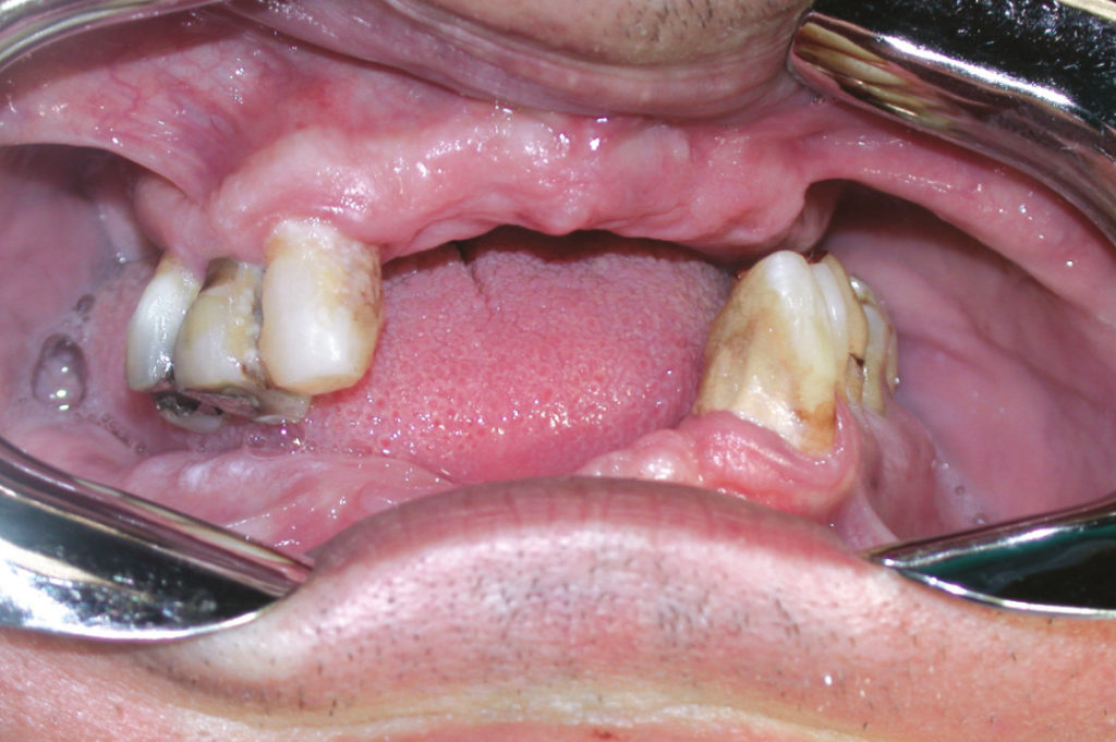
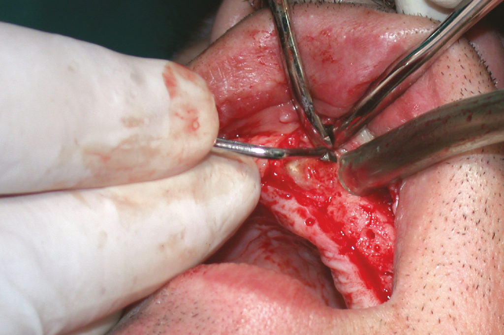
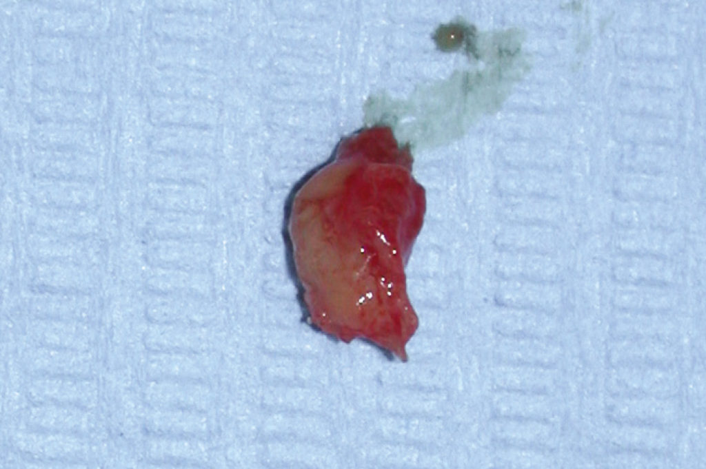
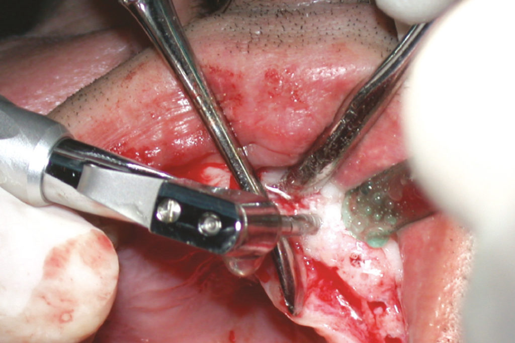
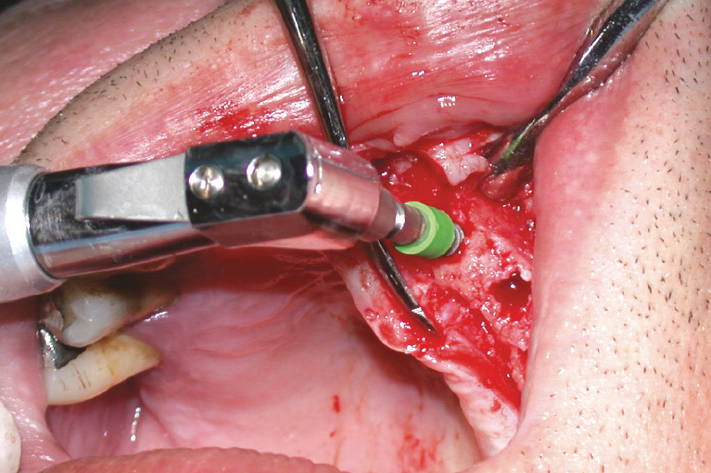
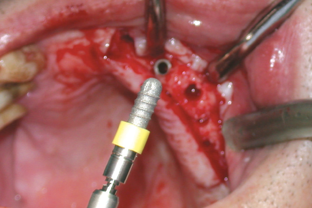
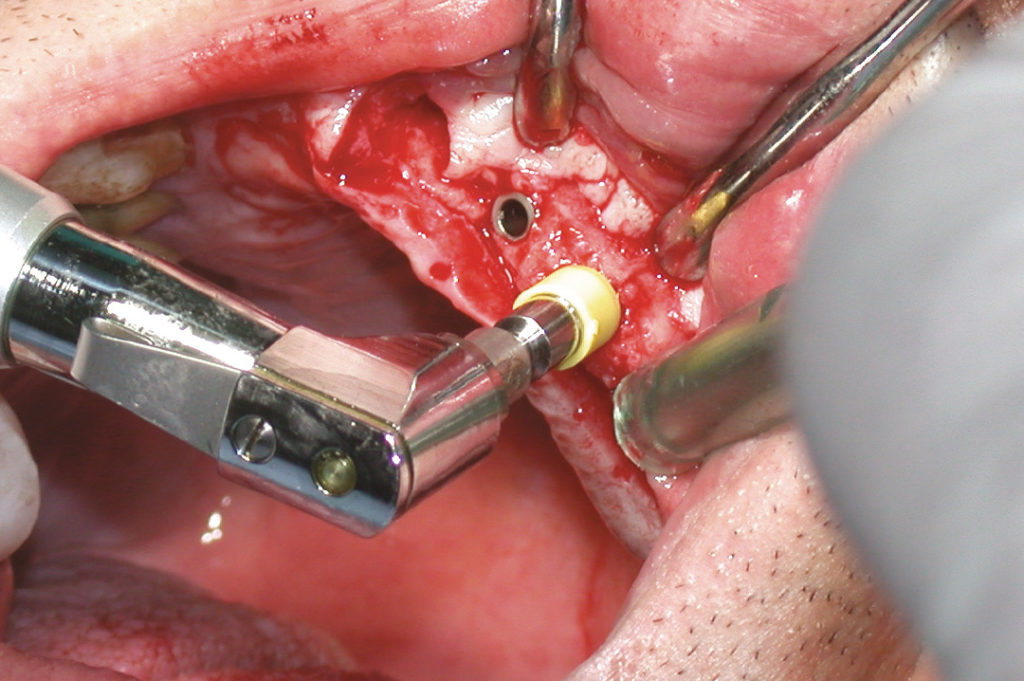
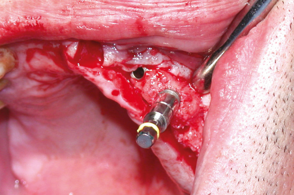
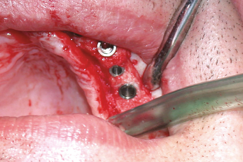
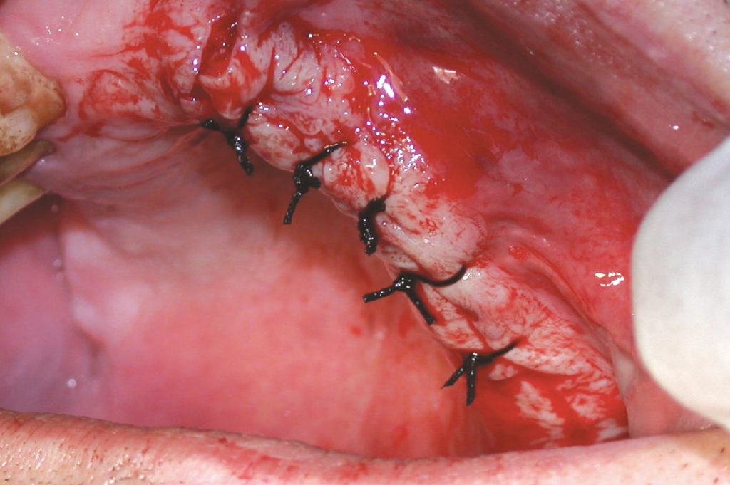
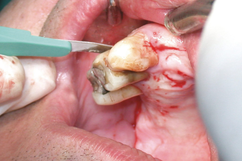
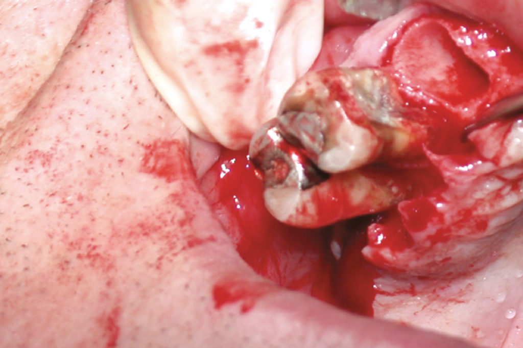
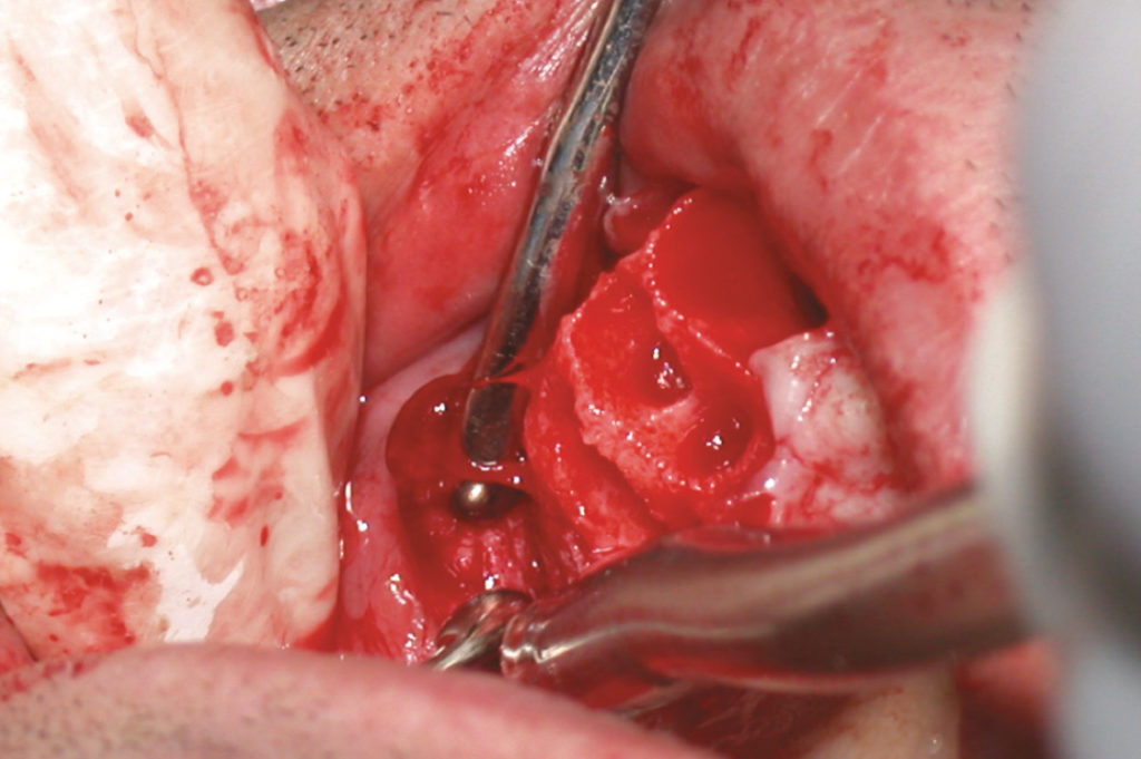
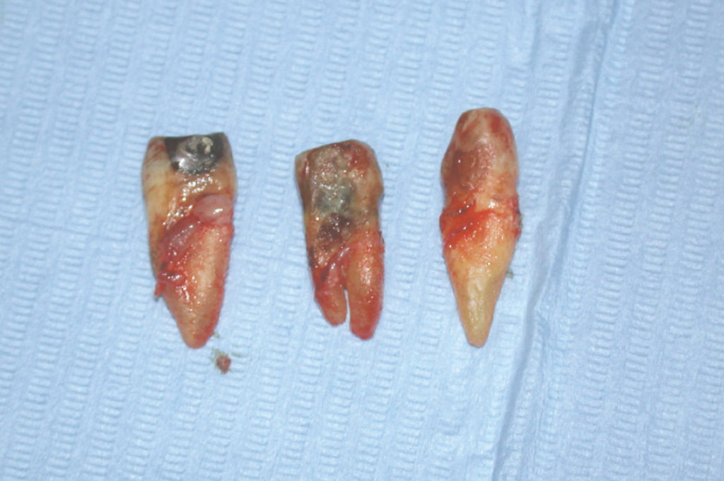
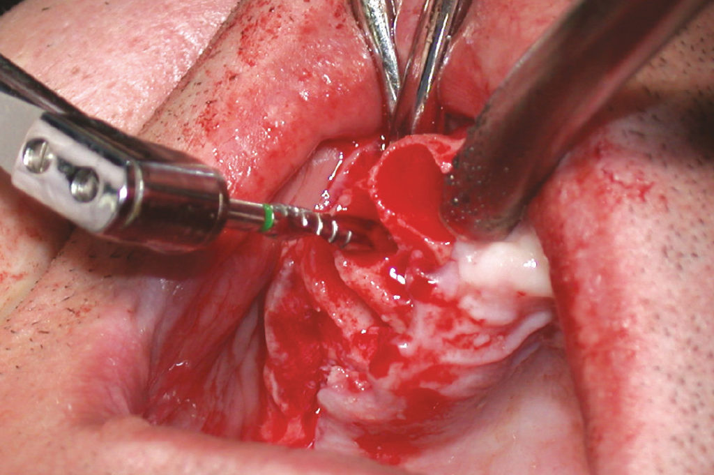
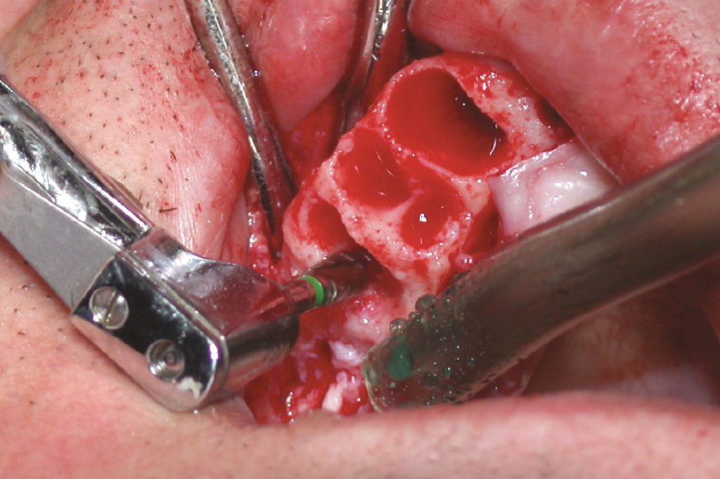
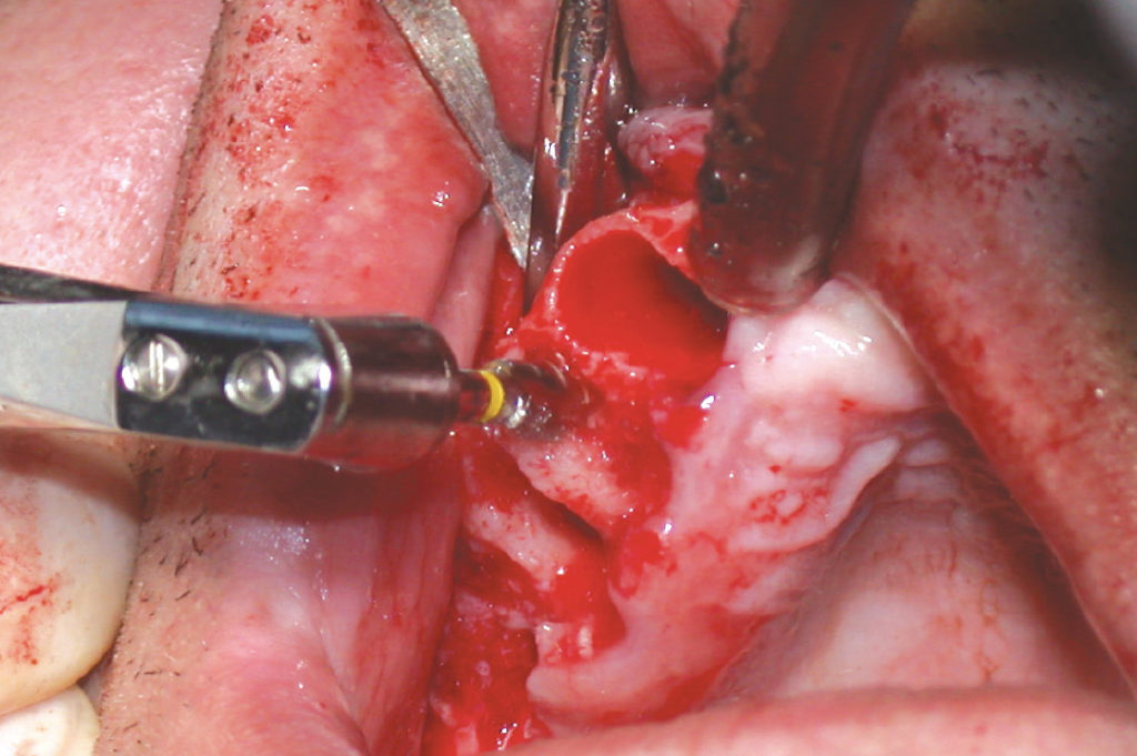
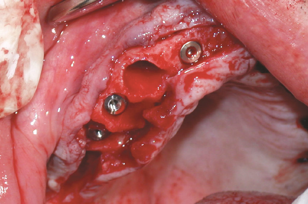
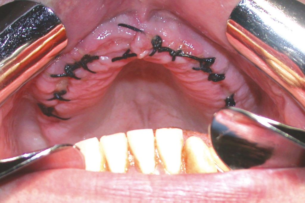
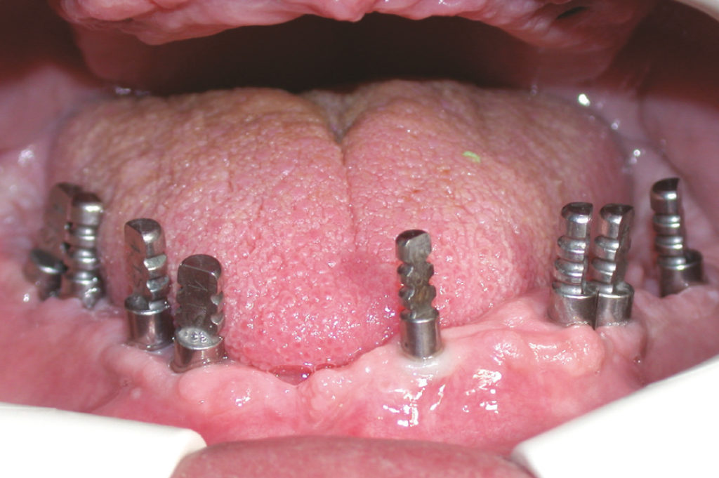
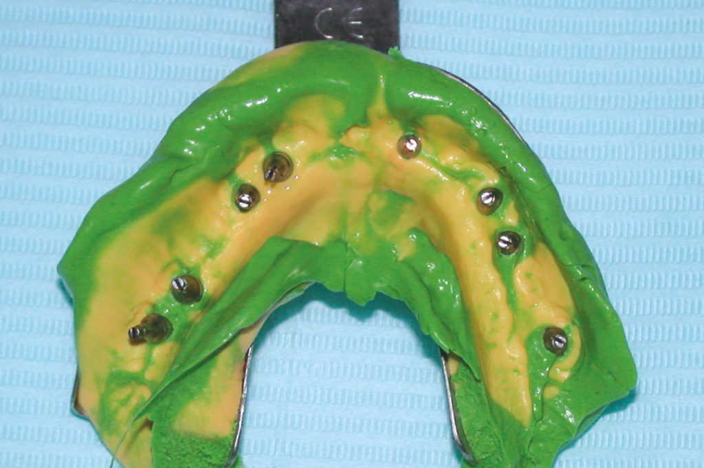
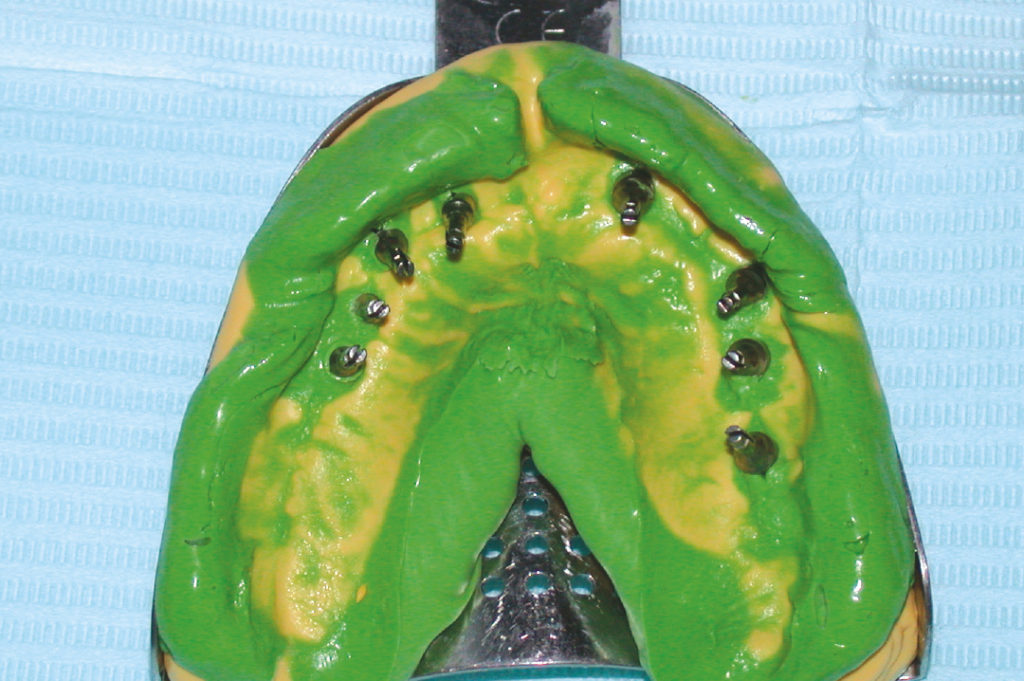
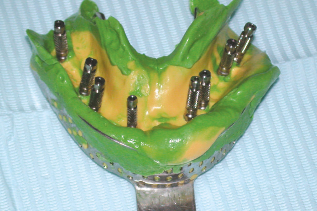
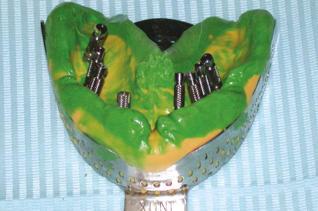
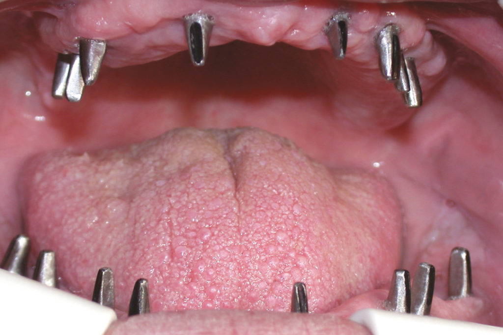
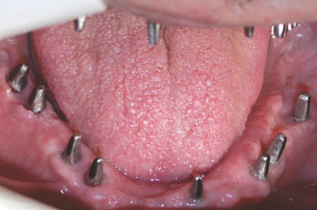
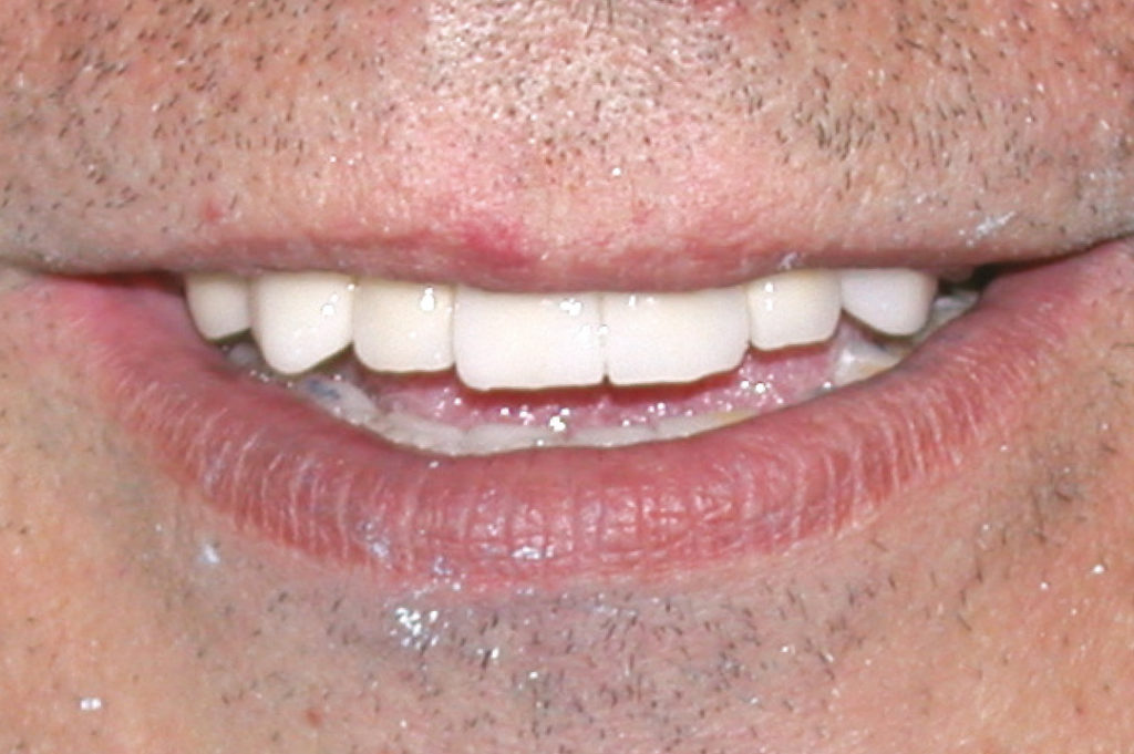
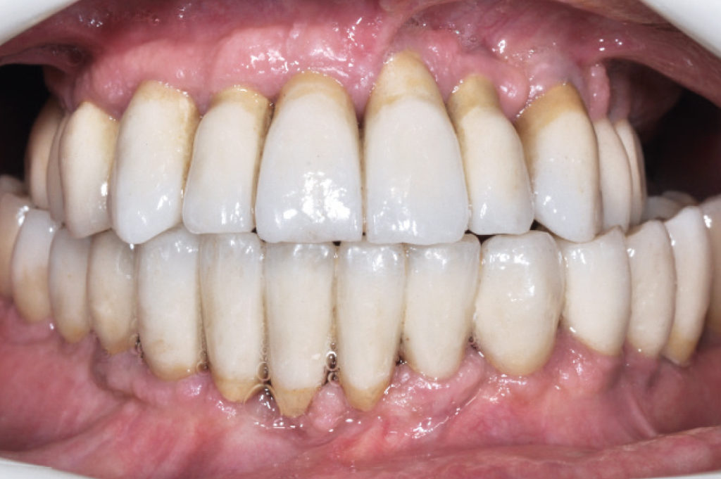
Pre-operative panoramic X-ray Pre-operative clinical view Cystectomy View of removed cyst Upper left quadrant: implant site preparation Placement of a 3.3 XCN Classix implant with the contra-angle handpiece Placement of a 4.1 XCN Classix implant with the contra-angle handpiece Placement of a 4.1 XCN Classix implant with the contra-angle handpiece View of 4.1 carrier on the implant Clinical view of implants placed in the upper left quadrant Tension-free wound closure with sutures Upper right quadrant: soft tissue incision Extraction of teeth #13, #14 and #15 Clinical appearance of the surgical site after tooth extraction Extracted teeth Upper right quadrant: implant site preparation Upper right quadrant: implant site preparation Upper right quadrant: implant site preparation Clinical appearance of upper right quadrant after placement of four implants View of upper jaw after placement of eight implants with a two-stage surgical procedure Clinical appearance of lower right quadrant after placement of four implants Clinical appearance of lower left quadrant after placement of four implants Panoramic X-ray six months after surgery prior to uncovering the implants View of transfers positioned in the upper implants for impression taking View of transfers positioned in the lower implants for impression taking Transfers inside the impression of the lower jaw Transfers inside the impression of the upper jaw Implant analogs fixed on the transfers inside the impression of the lower jaw Implant analogs fixed on the transfers inside the impression of the upper jaw Intra-oral try-in of the prepared abutments Intra-oral try-in of the prepared abutments Intra-oral try-in of the metal structures Patient’s smile after delivery of prostheses Follow-up panoramic x-ray after twelve years Clinical situation twelve years after delivery of the prosthesis showing stable gingival margins Comparison of 6-month follow-up and 12-year follow-up: the peri-implant marginal bone levels are stable
Laboratory:
Wilocs – Rome, Italy