Surgeons/Restorative dentists:
Dr. Giuseppe Musiello – Vico del Gargano (FG), Italy
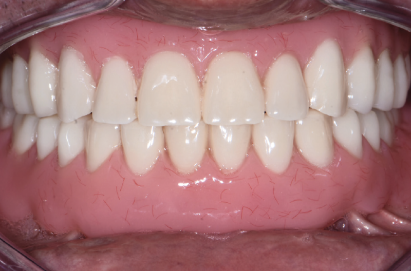
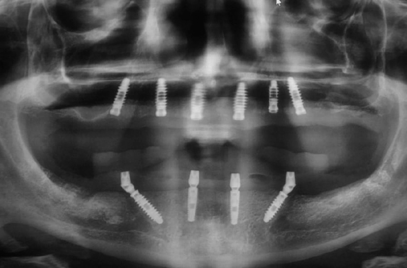
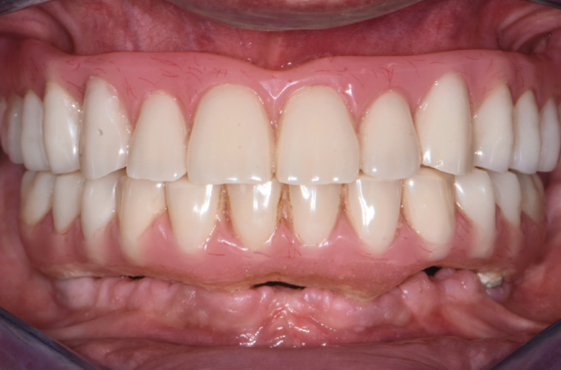
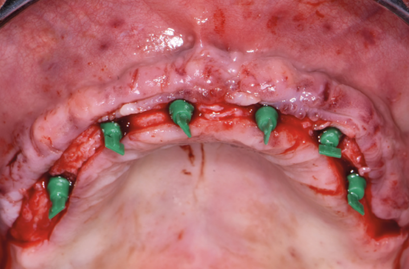
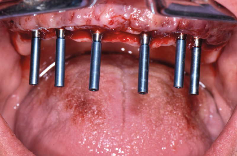
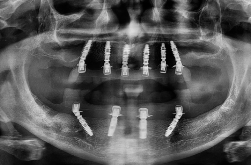
This case report shows the immediate loading of four XCN® implants in the lower jaw with a provisional fixed full-arch conometric prosthesis and the chairside fabrication of a provisional fixed full-arch conometric prosthesis on six osseointegrated implants in the upper jaw during the second surgical stage (re-opening). The 50-year-old completely edentulous male patient asked for a fixed restoration for both jaws. After careful study of CBCT examination and stabilized intermaxillary relationships with new diagnostic removable prostheses, it was decided to place ten implants, six in the maxilla and four in the mandible. As all four implants in the mandible had a good primary stability, implants were immediately provisionalized with a fixed conometric prosthesis. Due to the poor quantity and quality of bone, in the maxilla the immediate denture was inserted during the second surgical stage after 4 months of submerged healing. After 2 months, both provisional conometric prostheses were removed for impression taking on conometric abutments in order to fabricate two final fixed conometric prostheses with carbon fiber framework and the esthetic part with teeth and gingival composite portion.
XCN® conometric prosthesis obtains the friction retention through titanium abutments (MUA-Conics) with a 5-degree tapered top (half angle) and preformed conometric caps, made of PEEK, with an internal connection with the same taper angle, that are fixed within the prosthesis. The use of conometric friction retention as anchorage system for the prosthesis simplifies and speeds up all sessions. The ease with which a removable prosthesis can be transformed into a provisional fixed conometric prosthesis is a great advantage, especially in immediate loading. The speed with which conometric prostheses are removed and repositioned during all sessions (relining, impression taking, delivery of the final prosthesis) is one of the great advantages of this prosthesis type.

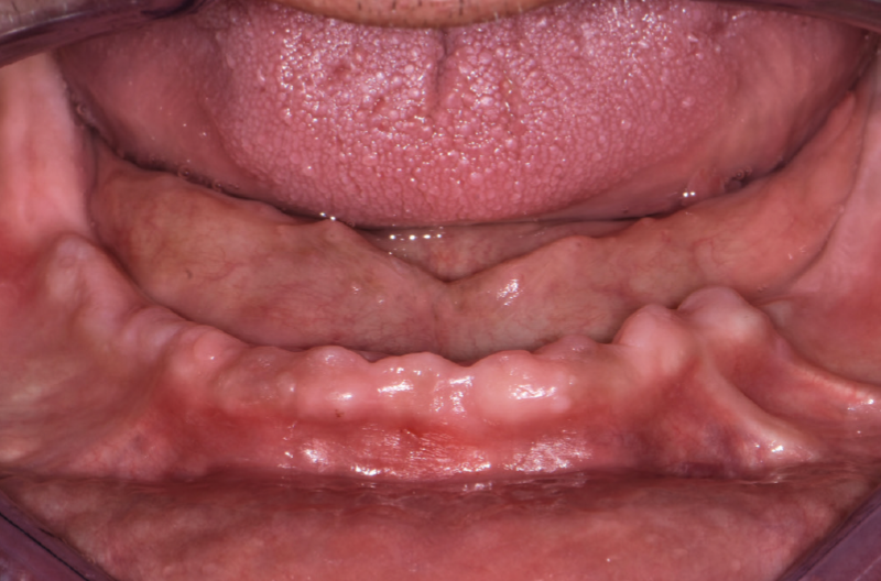
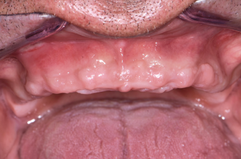
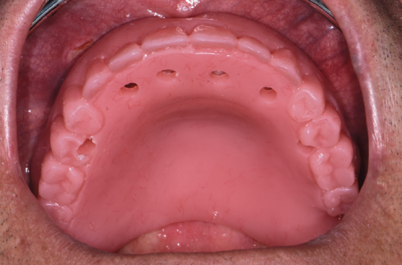
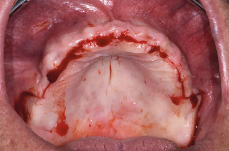
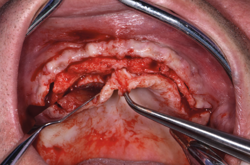
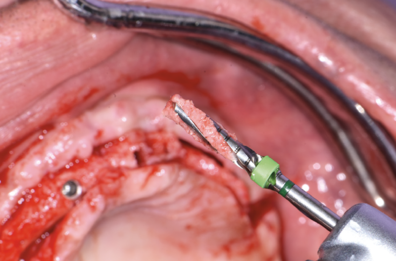
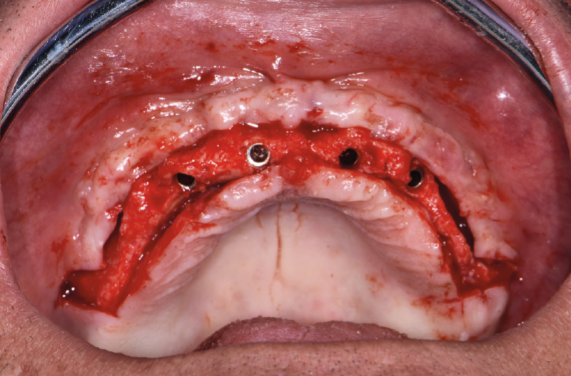
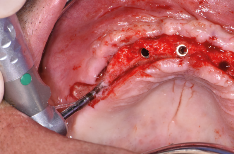
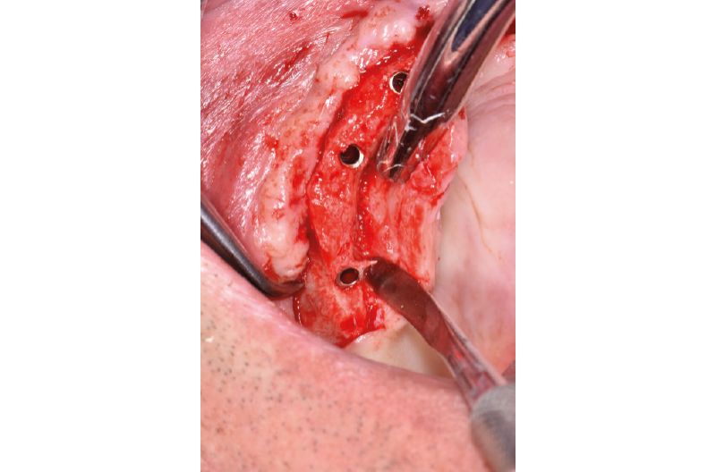
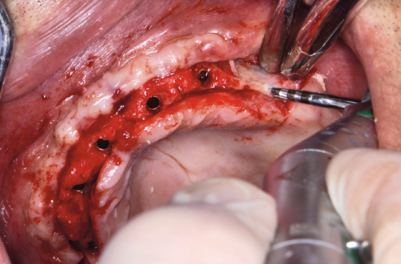
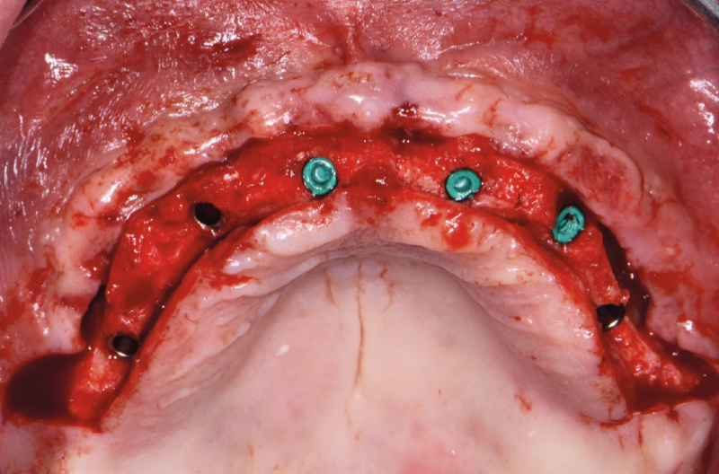
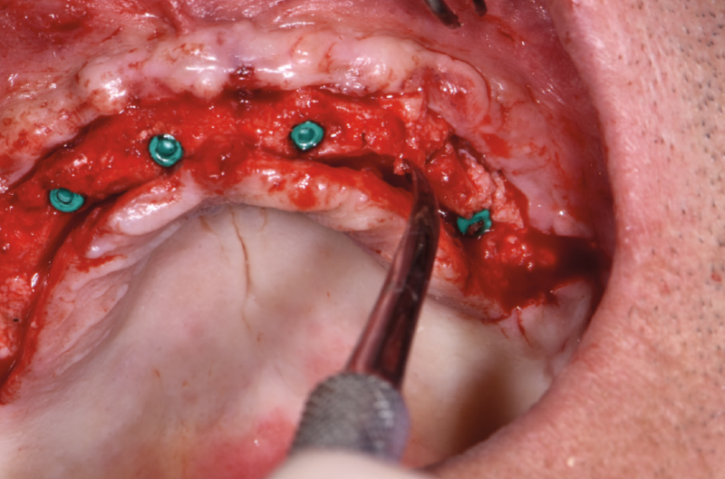
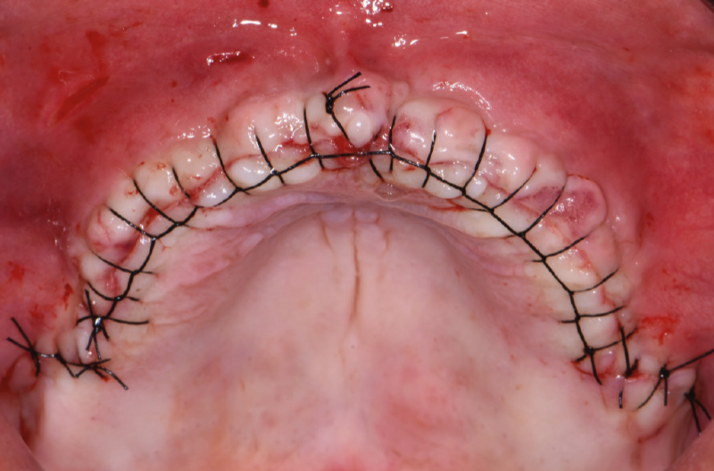
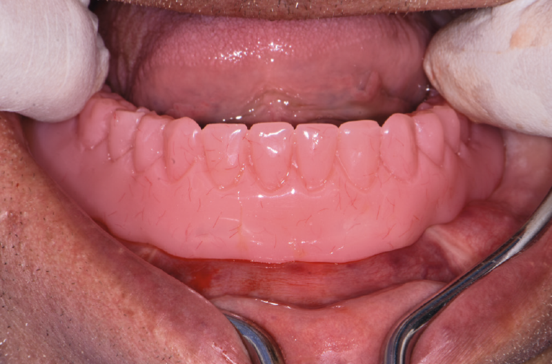
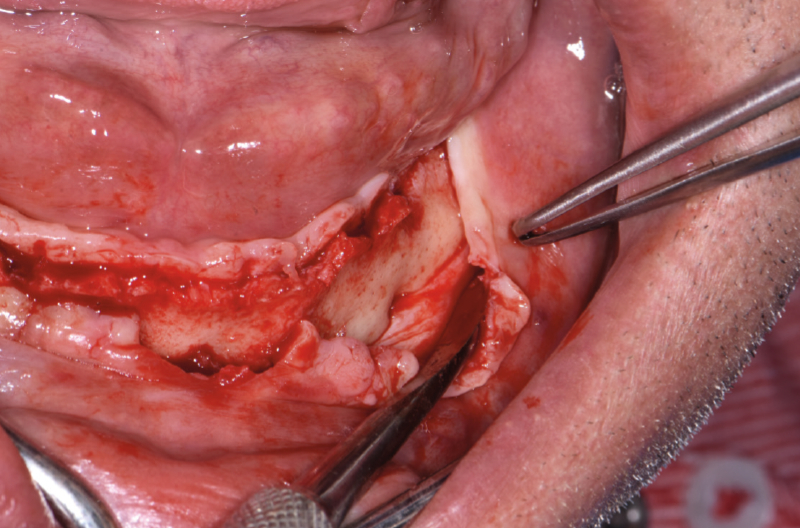
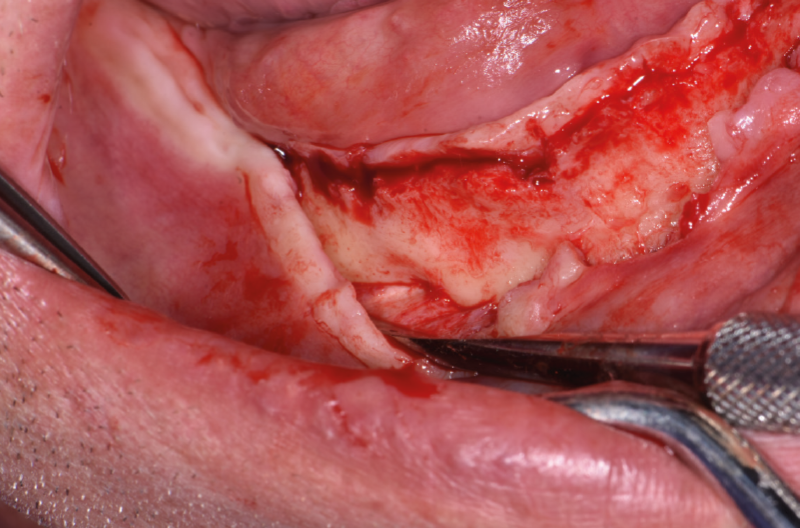
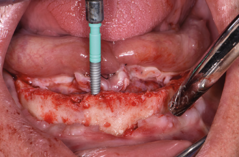
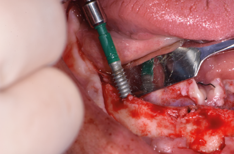
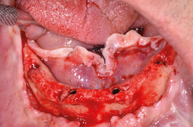
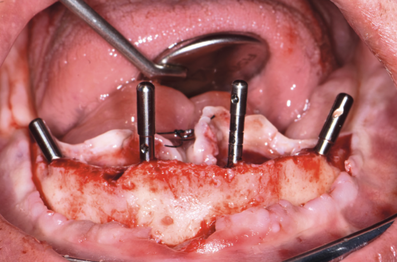
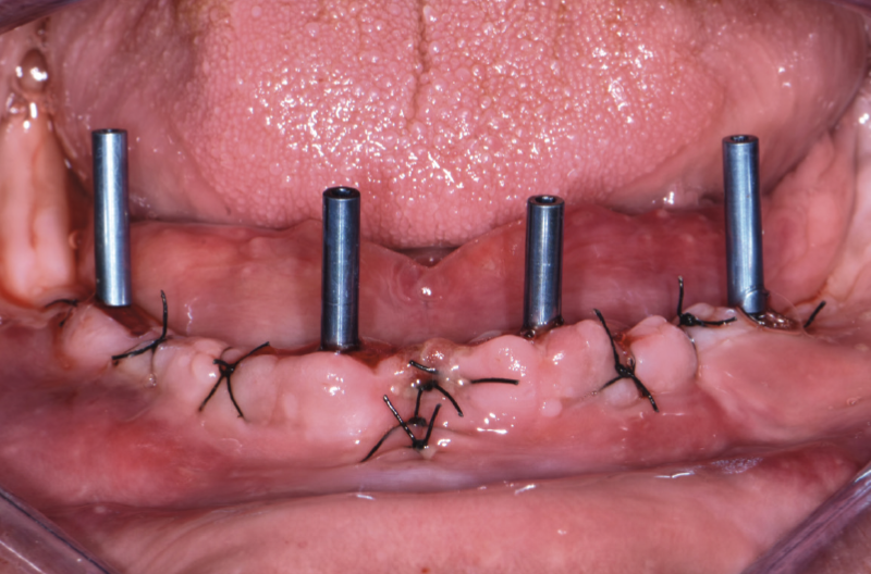
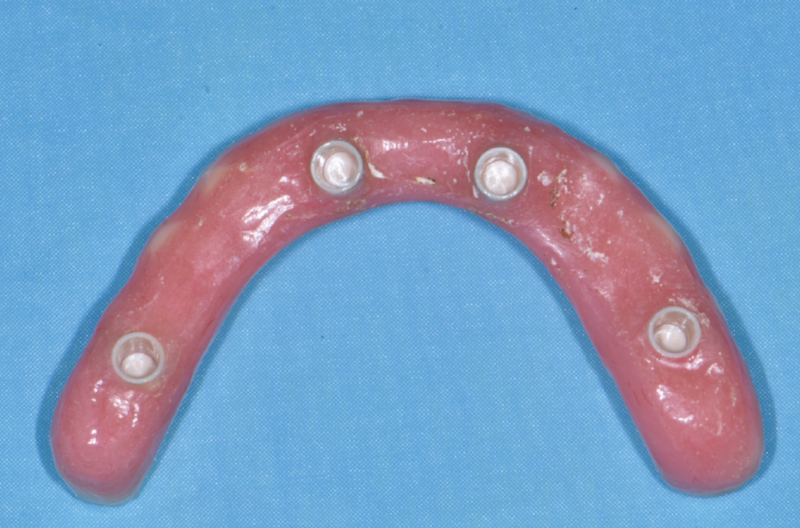
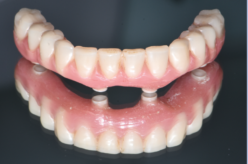
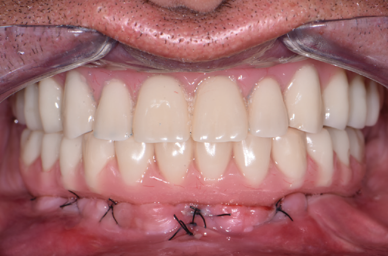
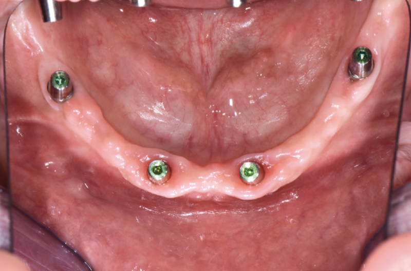
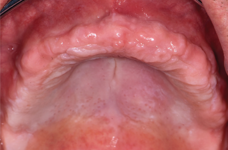
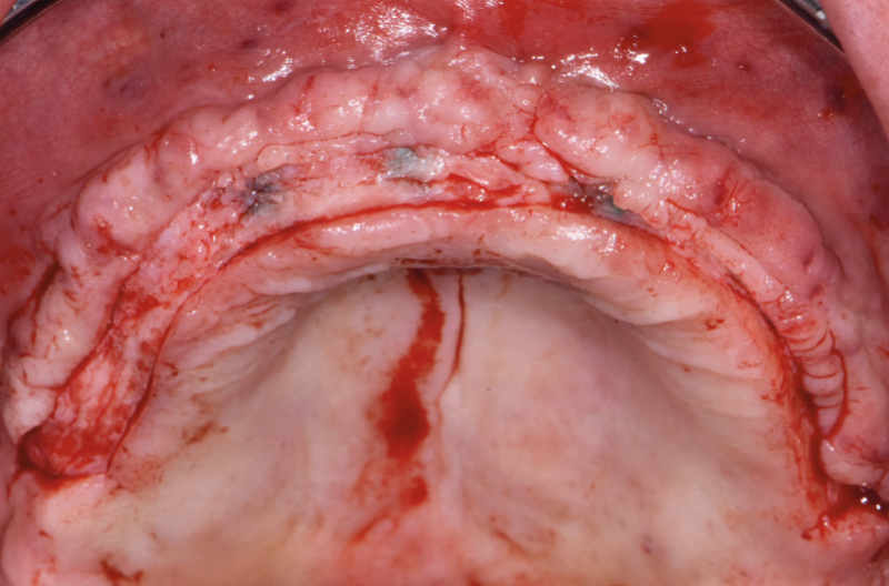
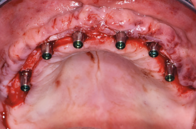
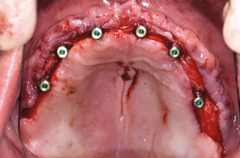
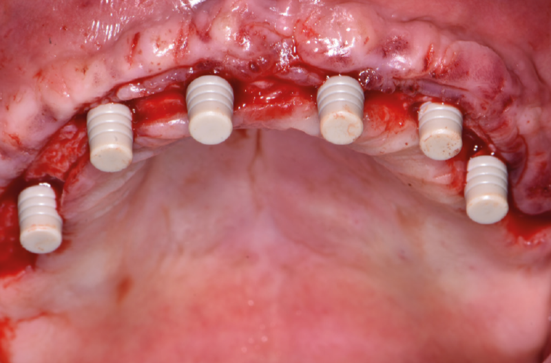
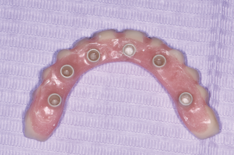
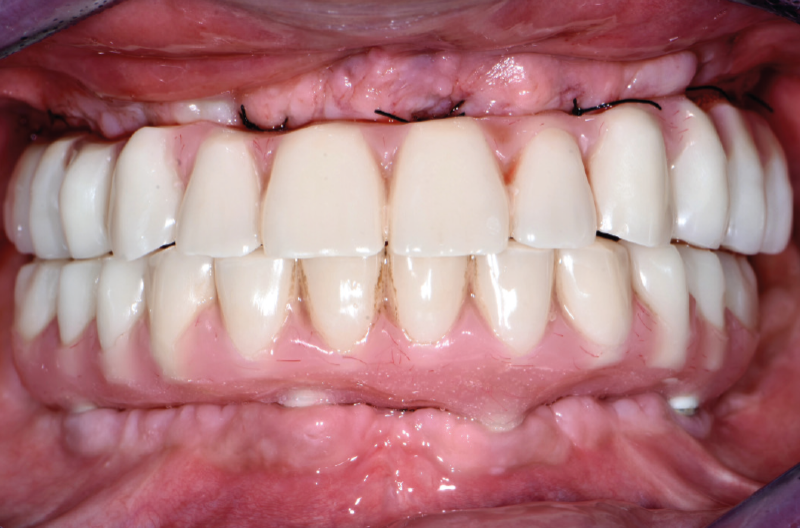
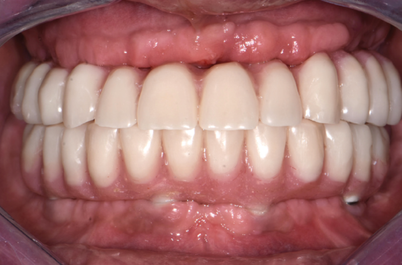
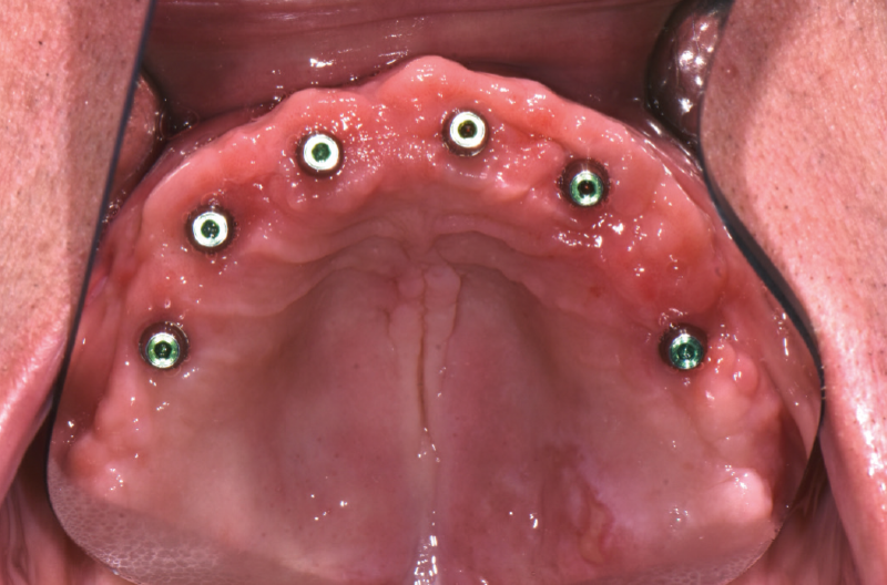
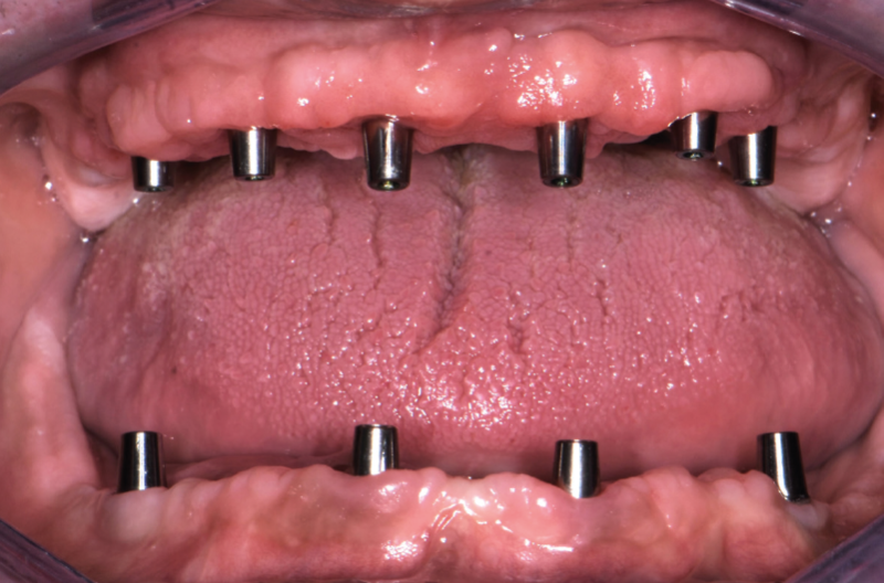
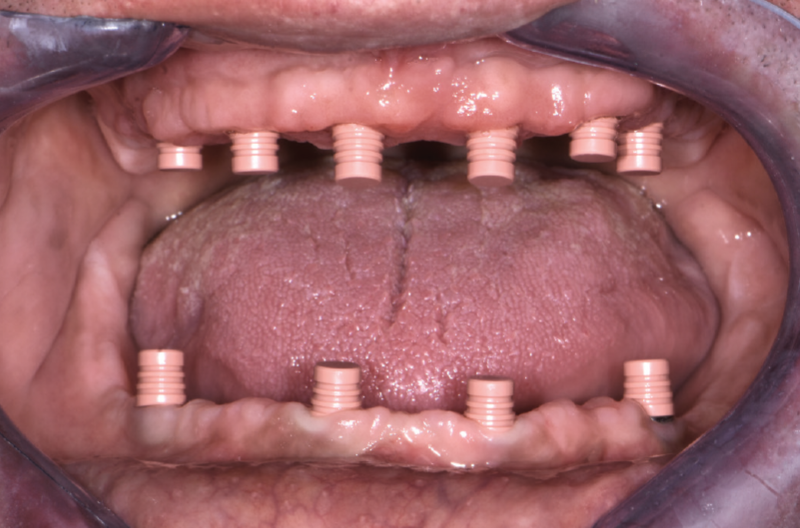
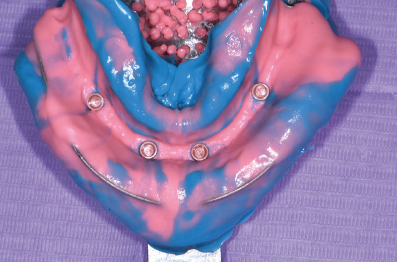
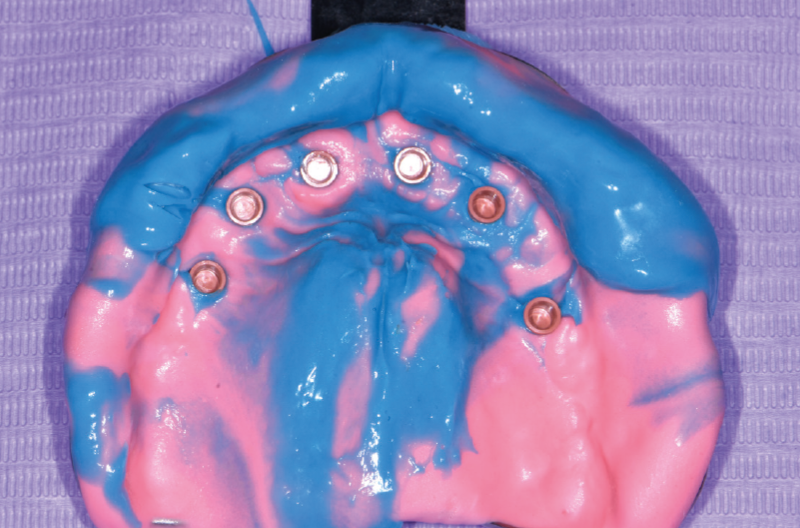
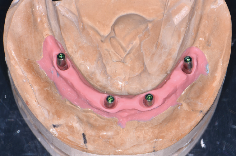
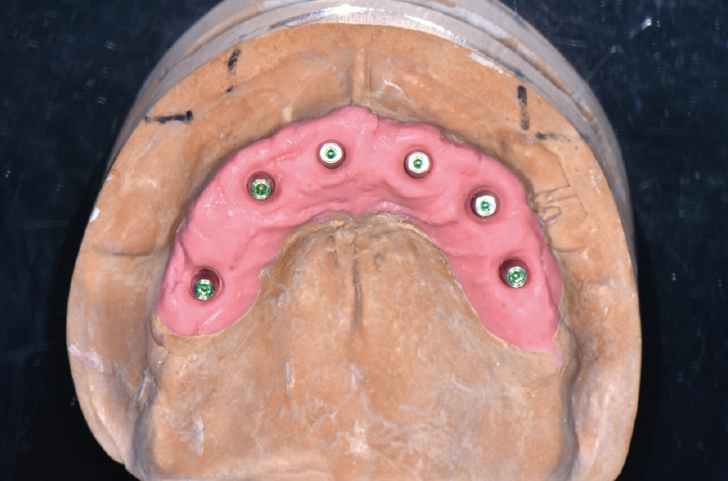
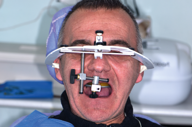
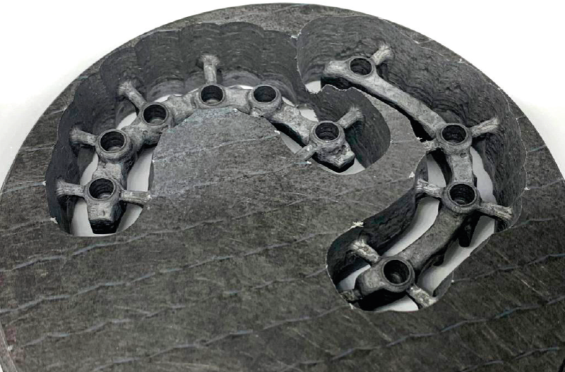
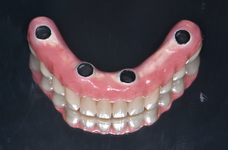
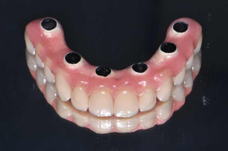
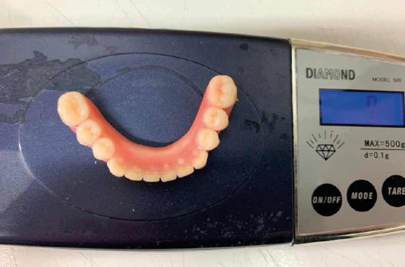
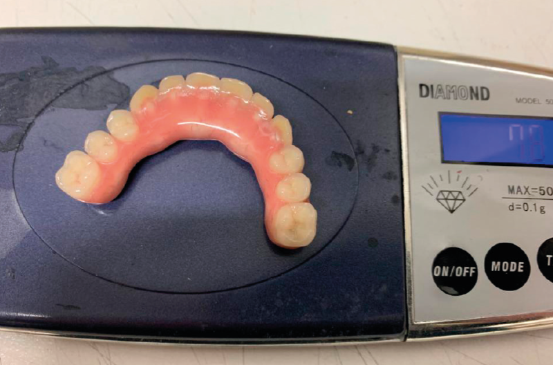
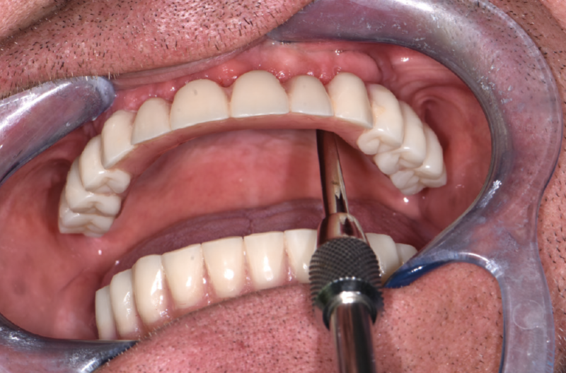
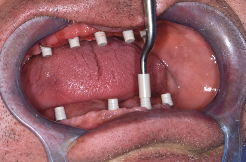
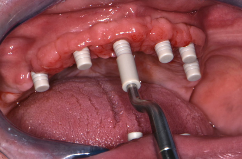
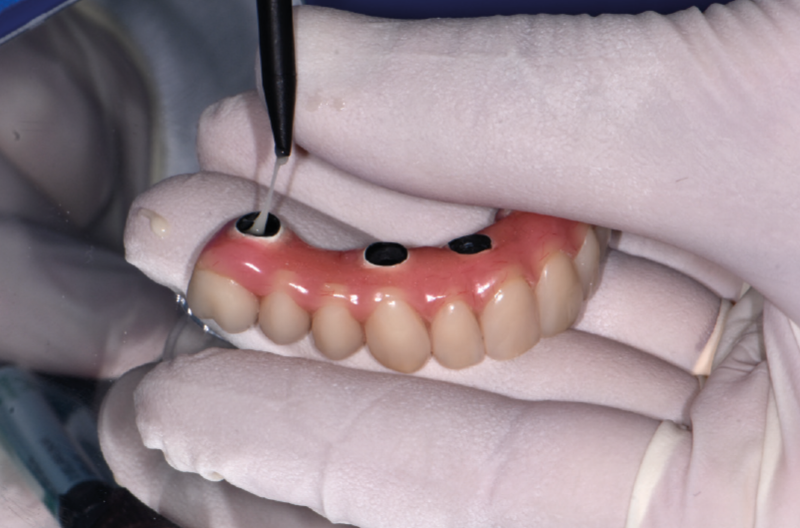
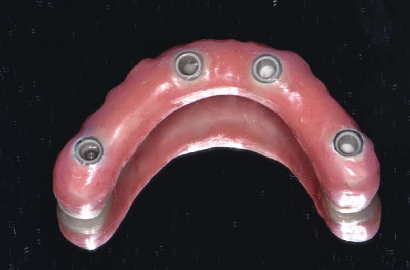
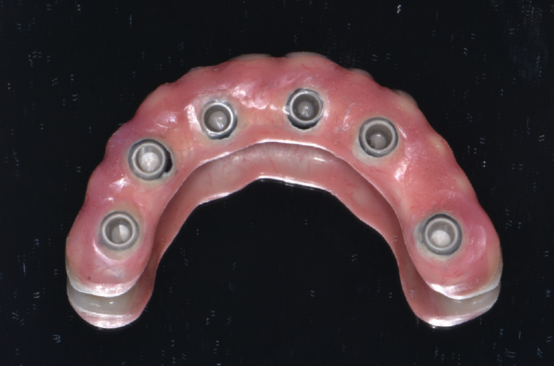
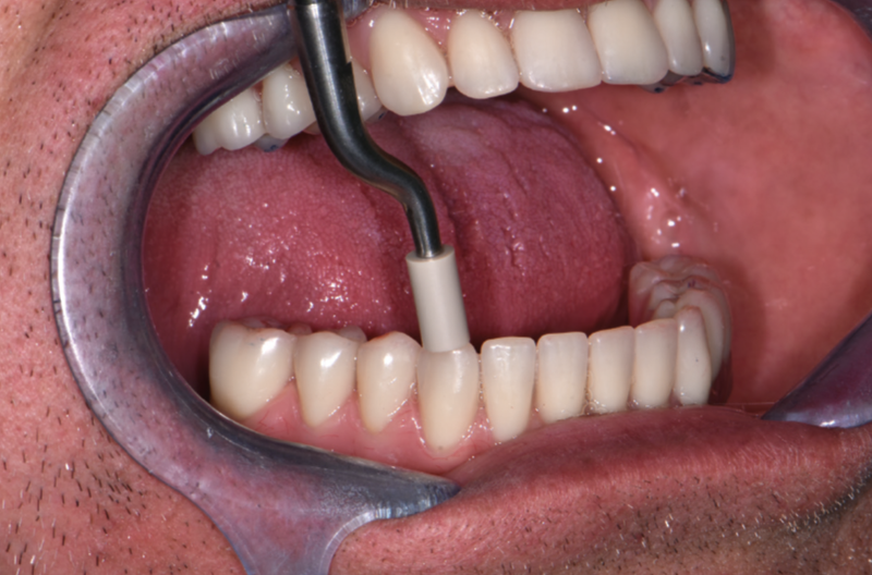
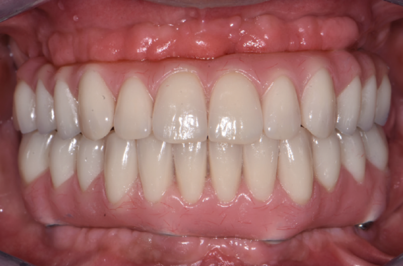
Initial removable prostheses View of clinical situation in the lower jaw View of clinical situation in the upper jaw Upper denture replica guide to mark the implant positions Full thickness mucoperiosteal flap Isolation of palatine nerve Autogenous bone recovery from drill flutes Four 3.75 x 12 mm Max Stability implants placed in the anterior region Use of special osseodensification burs in region #15 to increase bone density during implant site preparation and to move the medial wall of the right maxillary sinus Tilted 3.75 x 12 mm Max Stability implant in region #15 placed along the medial wall of the right maxillary sinus Use of special osseodensification burs in region #25 to increase bone density during implant site preparation and to move the medial wall of the left maxillary sinus Tilted 3.75 x 12 mm Max Stability implant in region #25 placed along the medial wall of the left maxillary sinus; Insertion of cover caps Autologous bone grafting Suture Lower denture replica guide to mark the implant positions Exposure of left mental foramen Exposure of right mental foramen Placement of two 2.9 x 12 mm Narrow implants in the anterior region Placement of two tilted 3.75 mm Max Stability implants (12 and 14 mm) in front of the mental foramen View of implants placed in lower jaw View of implant inclinations Insertion of two 7.5° angled GH 1.5 mm MUAs and of two 35° angled GH 3 mm MUAs. MUAs are mounted on a blu multifunctional screw for positioning and parallelization. MUAs were properly activated in the implants and Conic Adapters were screwed on MUAs to convert into MUA-Conics View of finished provisional conometric prosthesis. Conometric caps were incorporated intraorally within the prosthesis transforming the removable prosthesis into a provisional fixed conometric prosthesis Provisional fixed conometric prosthesis Final situation after surgery: removable prosthesis in the upper jaw for submerged healing of the implants and immediate loading in the lower jaw with provisional fixed conometric prosthesis Panoramic X-ray 4 months after implant placement Clinical situation 4 months after implant placement View of MUA-Conics in the lower jaw View of upper jaw Reopening of upper jaw with partial thickness flap Try-in abutments placed in the implants; one straight, three 15° angled and two 25° angled MUAs, all GH 1.5 mm, were chosen Insertion and palallelization of MUAs; blu multifunctional screws were unscrewed and abutments properly tapped into the implants Conic Adapters were screwed on MUAs and tightened with the prosthetic torque wrench to convert into MUA-Conics Occlusal view of MUA-Conics in the upper jaw Fixed caps positioned onto MUA-Conics by performing 1 percussion with the PEEK tip View of finished provisional upper conometric prosthesis. Conometric caps were incorporated intraorally within the prosthesis with reline acrylic resin transforming the removable prosthesis into a provisional fixed conometric prosthesis View of upper and lower provisional conometric prostheses in place After 1 month, removal of both provisional conometric prostheses and relining for better soft tissue conditioning Occlusal view of clinical situation after removal of the provisional conometric upper prosthesis 1 month later Clinical situation at time of impression taking on MUA-Conics Fig. 41 Mobile caps used as transfer copings for the impression taking on MUA-Conics Lower jaw impression Upper jaw impression Dental cast of the lower jaw prepared with MUA analogs and Conic Adapters Dental cast of the upper jaw prepared with MUA analogs and Conic Adapters Registration of the vertical dimension Fabrication of carbon fiber frameworks for the final prostheses Ridge-side view of lower final prosthesis: note appropriate spaces for intraoral cementation of Fixed caps into the prosthesis Ridge-side view of upper final prosthesis: note appropriate spaces for intraoral cementation of Fixed caps into the prosthesis Final lower prosthesis with carbon fiber framework weighs only 9.1 gr Final upper prosthesis with carbon fiber framework weighs only 7.8 gr Removal of provisional conometric prostheses by gentle tapping force applied laterally on the bridge with PEEK tip and use of crown remover Fixed caps positioned onto MUA-Conics in the lower jaw by performing 1 percussion with the PEEK tip Fixed caps positioned onto MUA-Conics in the upper jaw by performing 1 percussion with the PEEK tip Intraoral cementation of final prostheses on Fixed caps with a dual resin cement View of final lower prosthesis with incorporated Fixed caps after excess cement cleaning and polishing View of final upper prosthesis with incorporated Fixed caps after excess cement cleaning and polishing Activation of final prostheses on MUA Conics by tapping with the PEEK tip on the teeth nearby MUA-Conics View of final prostheses activated on MUA-Conics Panoramic X-ray after delivery of final prostheses. Note that Fixed caps, made of PEEK, are radiolucent
Laboratory:
Pasquale Martino – Foggia, Italy