Surgeon/Restorative dentist:
Dr. Renato Turrini – Massarosa (Lucca), Italy
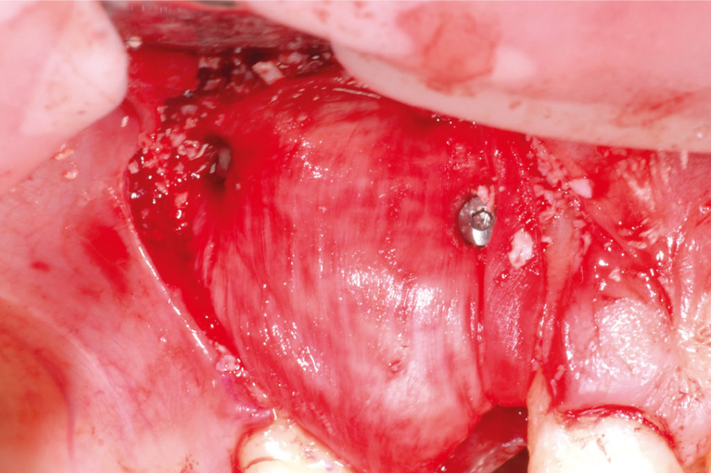
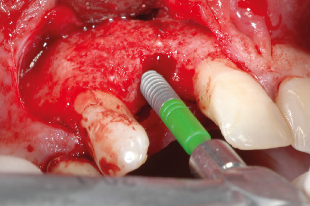
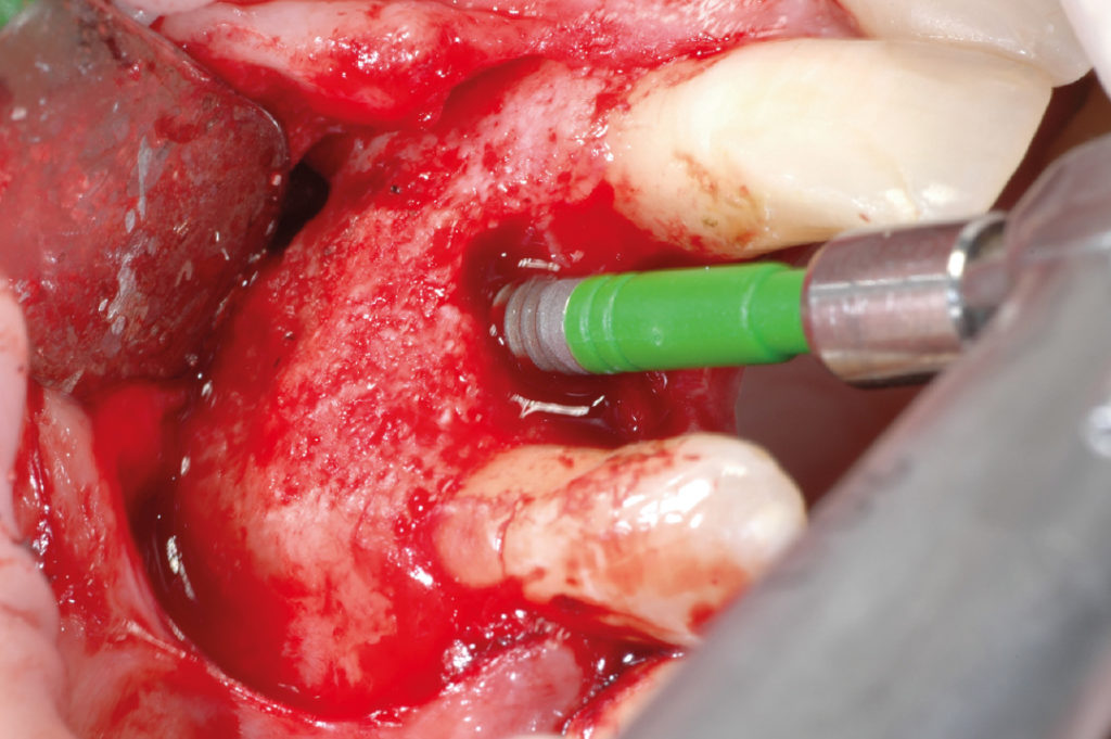
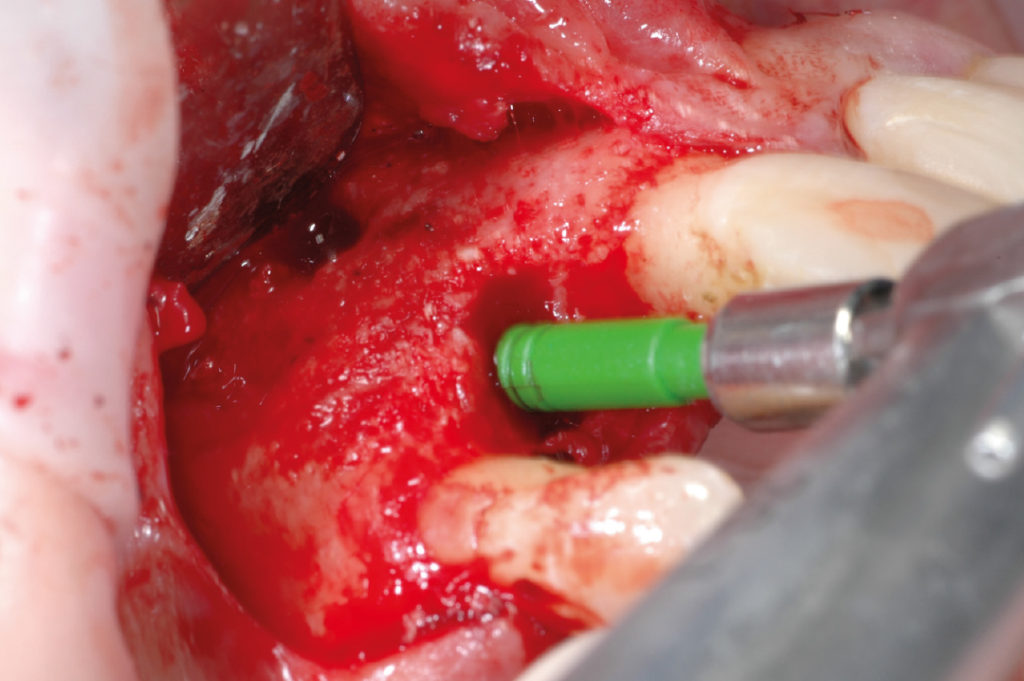
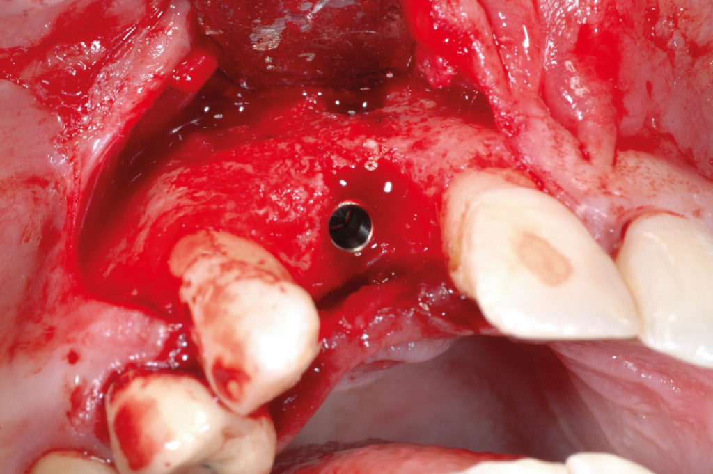
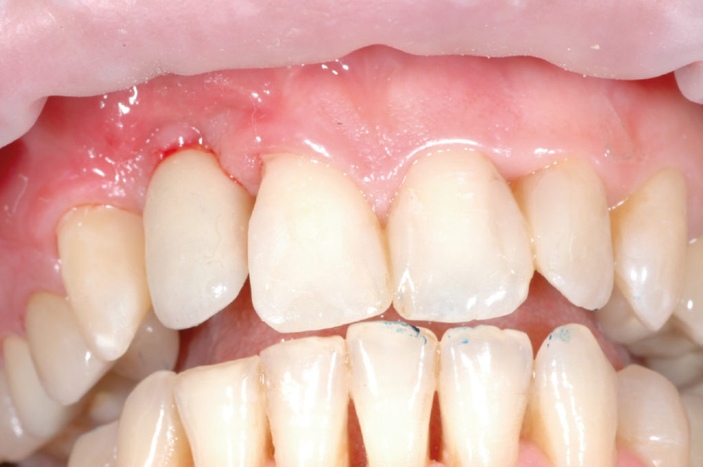
This case illustrates bone grafting with a combination of autologous bone graft and synthetic bone material covered by a membrane stabilized with tacks in order to regenerate the concave osseous architecture of the buccal bone plate. After six months of healing the fixation tacks were removed and a two-stage placement of a 2.9 x 12 mm XCN implant with simultaneous soft tissue thickening was performed. Three months later, the implant was uncovered, soft tissue was conditioned with a healing cap and an implant level impression was taken for the fabrication of a porcelain crown.

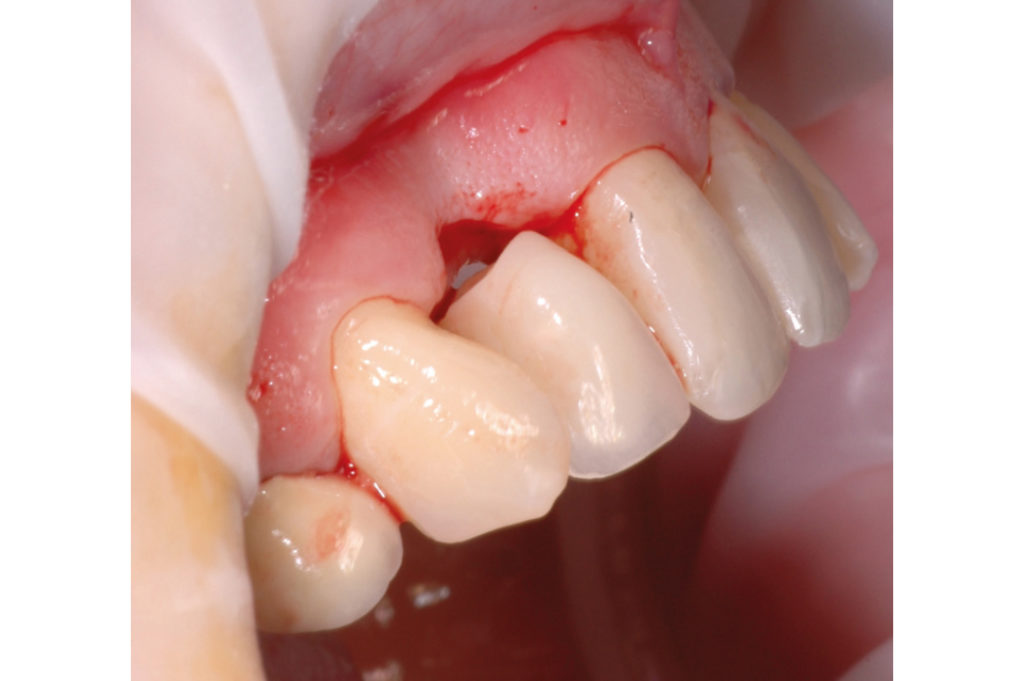
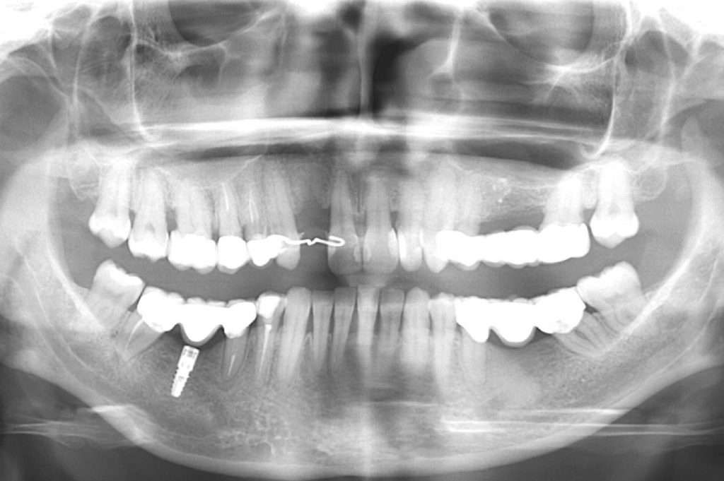
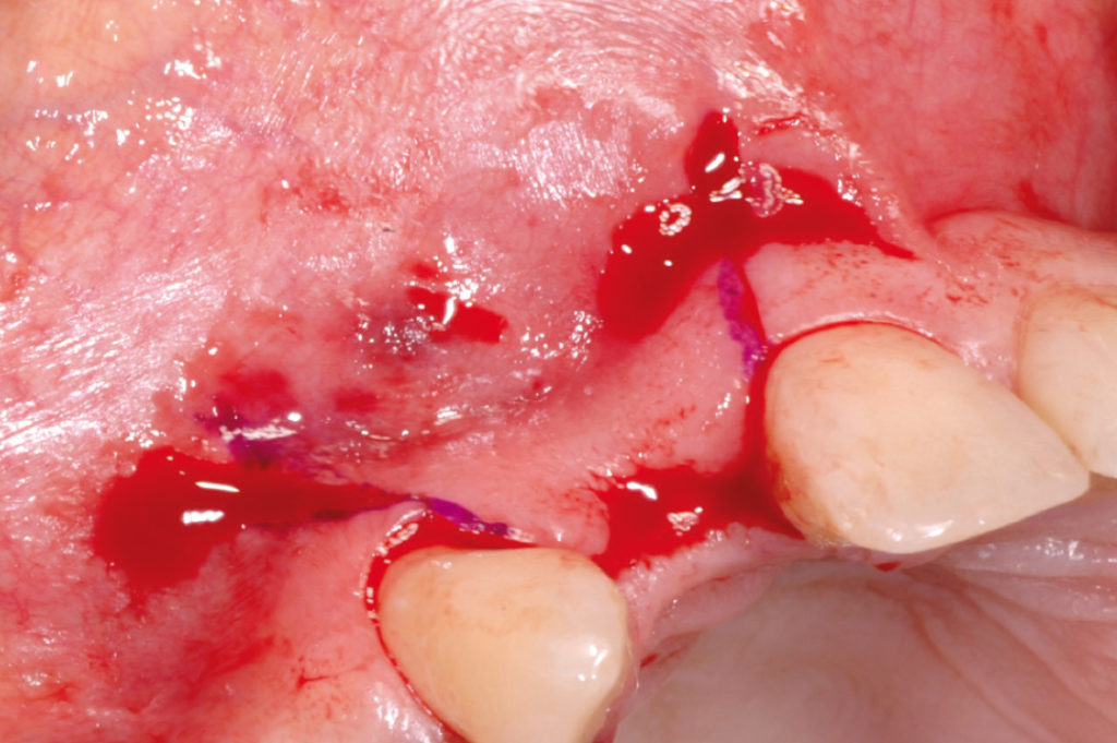
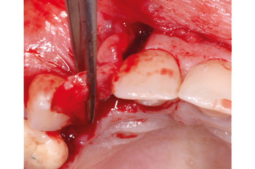
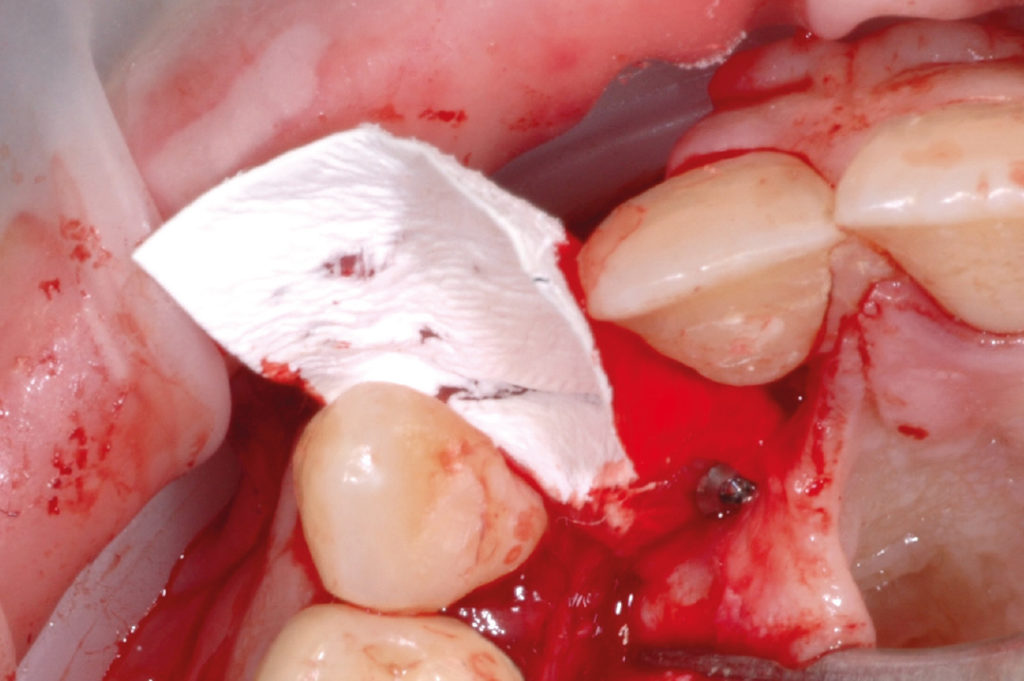
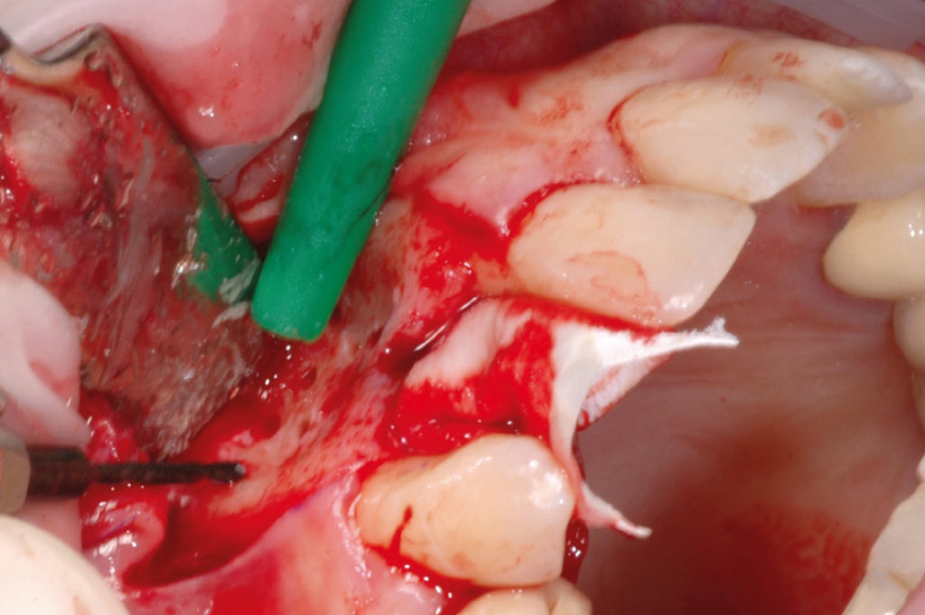
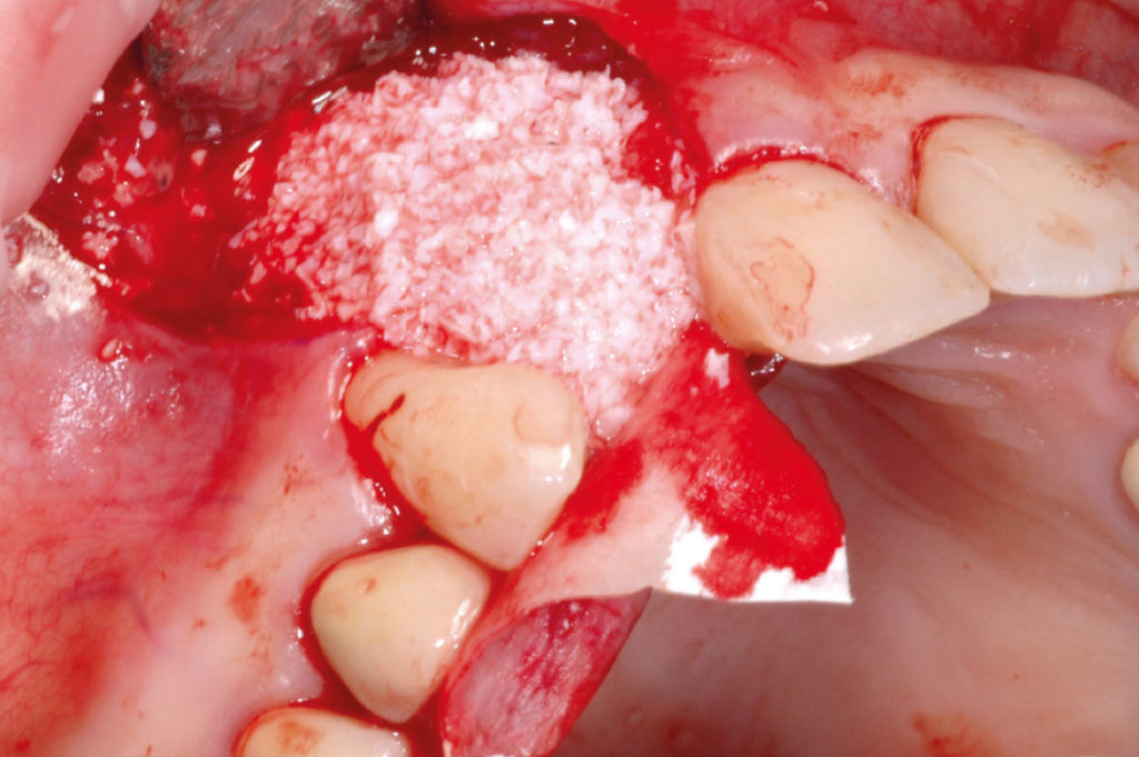
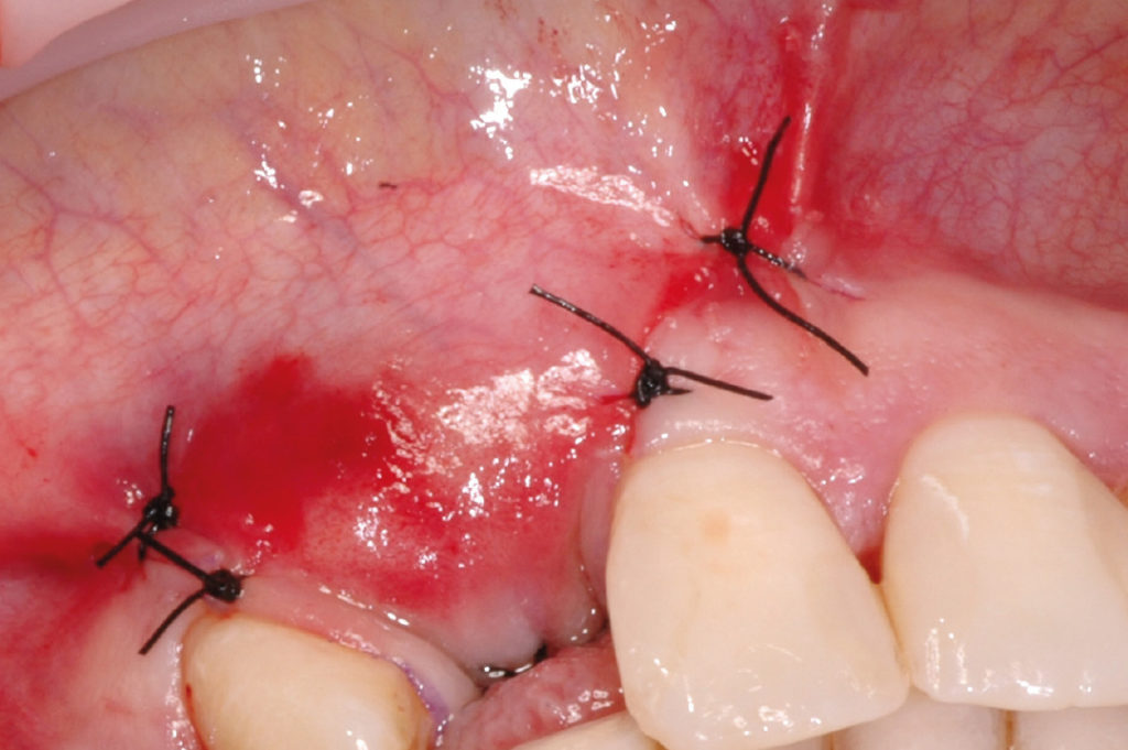
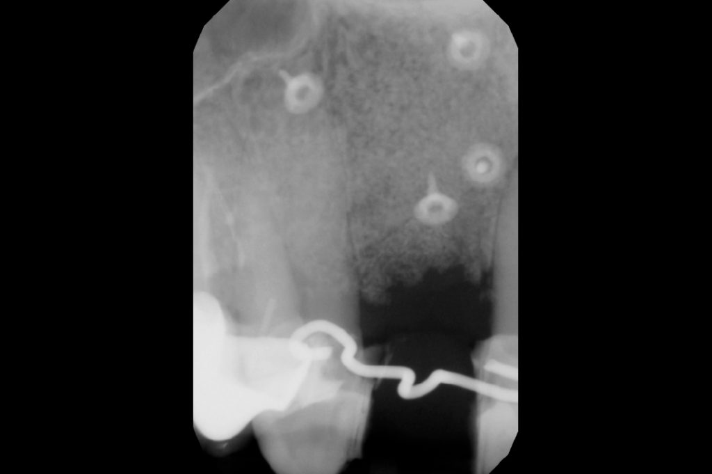
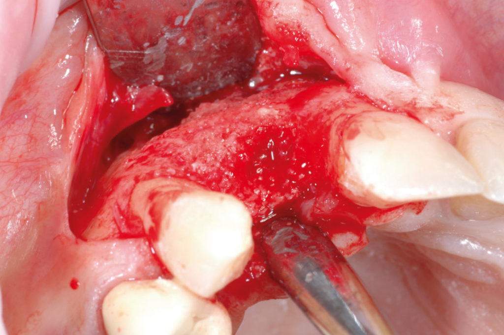
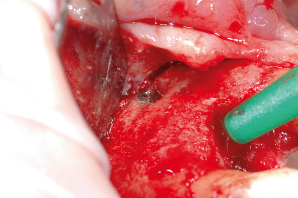
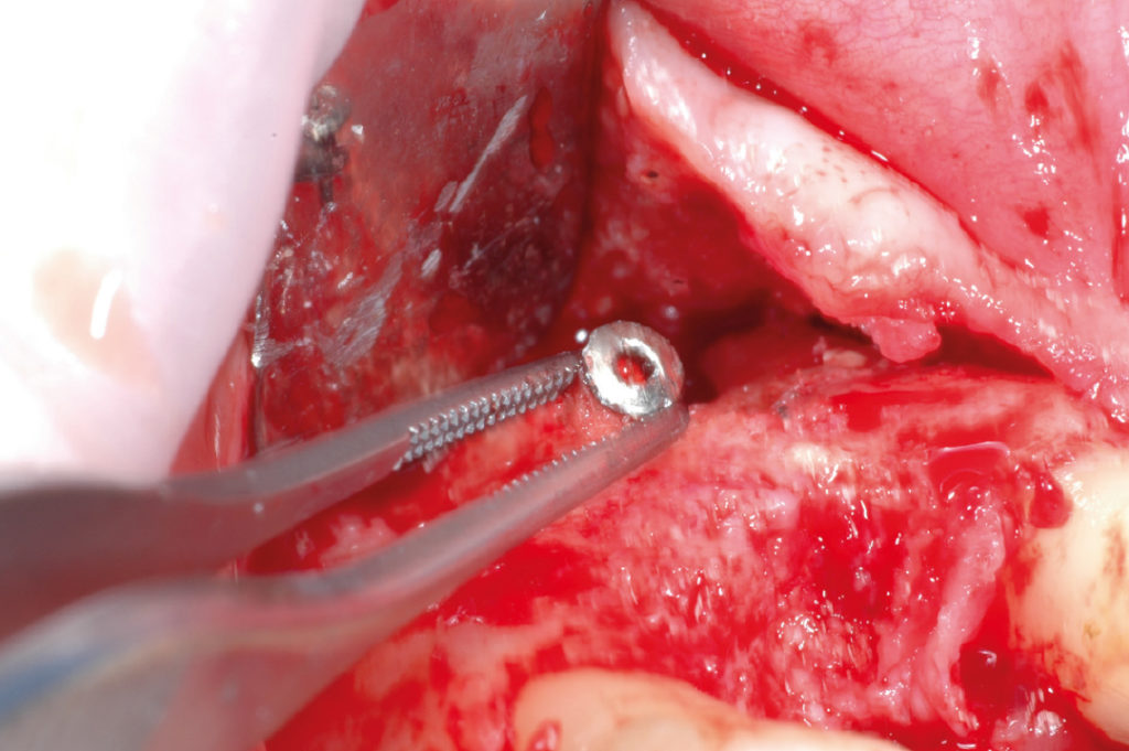
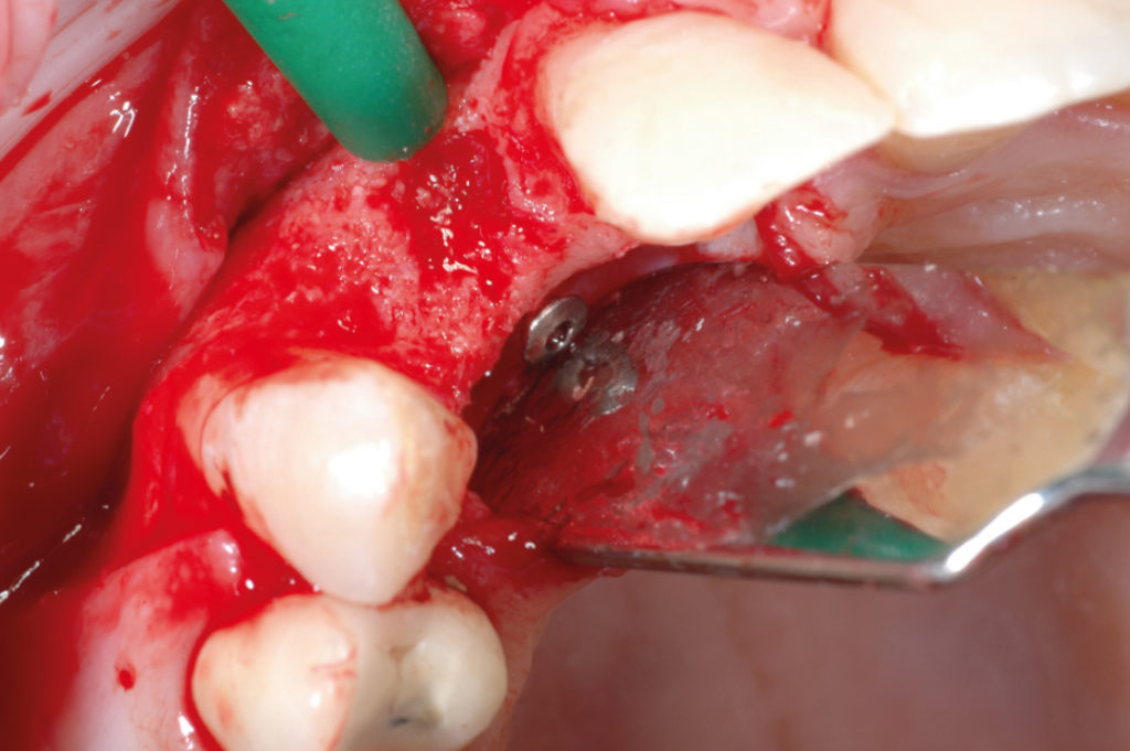
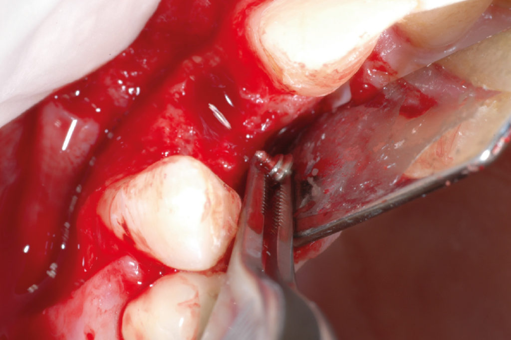
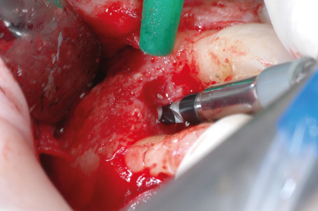
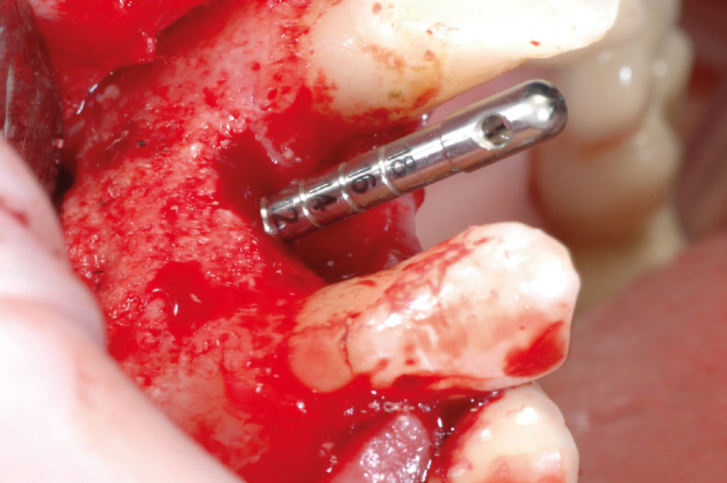
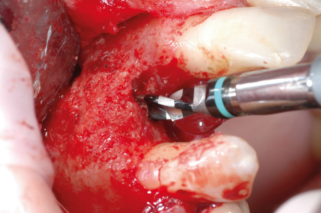
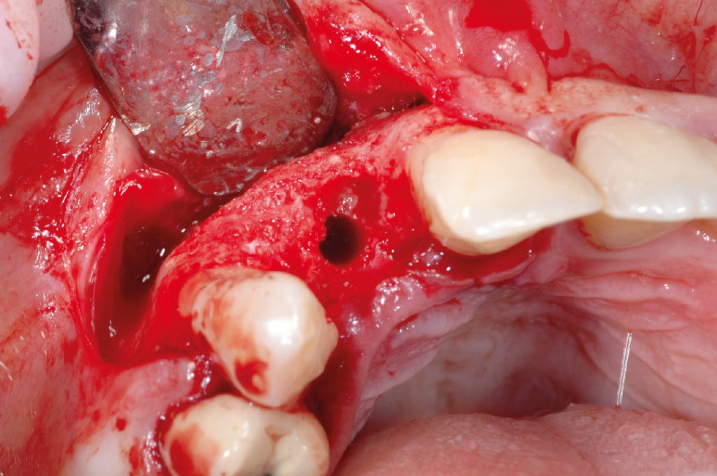
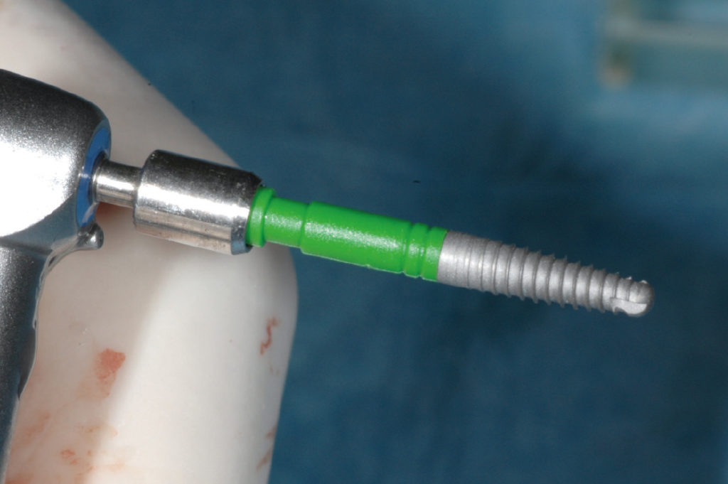
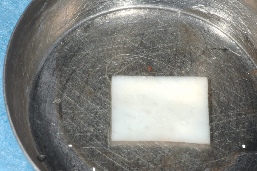
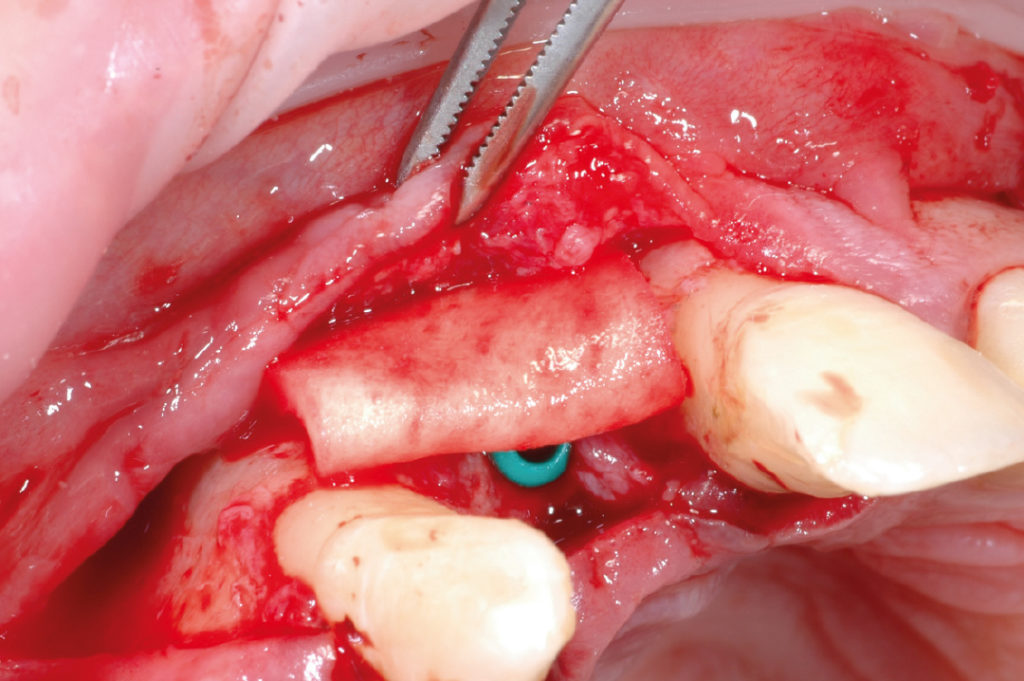
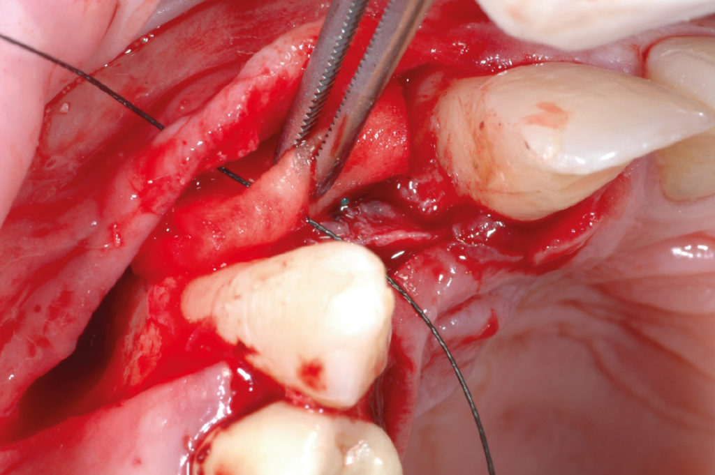
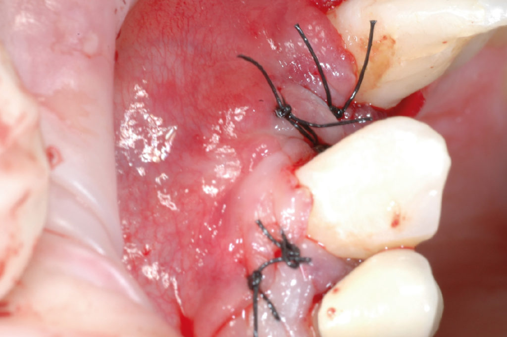
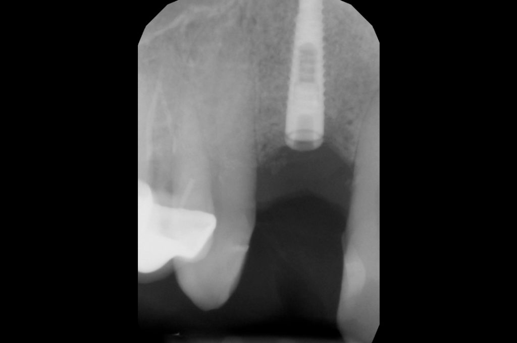
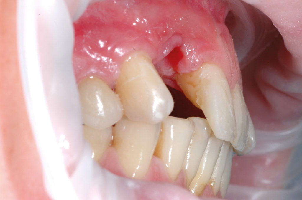
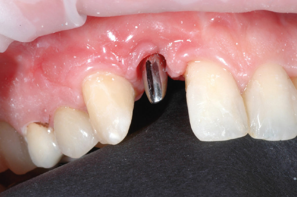
Pre-operative clinical view. Note the hard and soft tissue deficit in position #12 Pre-operative panoramic X-ray Full thickness flap with two vertical releasing incisions Flap reflection to expose the bone Membrane secured palatally by means of fixation tacks Small cortical perforations to increase blood supply Mixture of autologous bone graft harvested from the mandibular and synthetic bone material in place Membrane placed over bone graft and fixed with tacks Tension-free wound closure and suturing X-ray examination after six months Full thickness flap to expose the bone View of fixation tack on buccal plate Removal of buccal fixation tack View of fixation tack on palatal plate Removal of palatal fixation tack Use of 2.2 mm pilot drill for a length of 12 mm Use of 2.2 mm pilot drill for a length of 12 mm Depth gauge to check proper depth Use of 2.8 mm twist drill for a length of 6.5 mm to finalize site preparation View of created osteotomy 2.9 x 12 mm XCN implant coupled with is carrier connected to the contra-angle handpiece Implant insertion with contra-angle handpiece Implant insertion with contra-angle handpiece Implant insertion with contra-angle handpiece Implant in place before cover cap application for two-stage surgery Collagen-based tissue matrix Application of collagen-based tissue matrix for soft tissue thickening Suturing to secure the tissue matrix and to close the surgical wound Post-operative clinical view Post-operative X-ray Clinical view after soft tissue conditioning. Note the resolution of the hard and soft tissue deficiencies in position #12 Try-in of the titanium abutment Delivery of the porcelain crown

Laboratory:
Fratelli Fruzzetti – Viareggio, Italy